sc-PDB
An Annotated Database of Druggable Binding Sites from the Protein DataBank
An Annotated Database of Druggable Binding Sites from the Protein DataBank
1.950 Å
X-ray
2005-12-14
| Name: | Cytochrome P450 2A6 |
|---|---|
| ID: | CP2A6_HUMAN |
| AC: | P11509 |
| Organism: | Homo sapiens |
| Reign: | Eukaryota |
| TaxID: | 9606 |
| EC Number: | 1.14.13 |
| Chain Name: | Percentage of Residues within binding site |
|---|---|
| A | 100 % |
| B-Factor: | 20.157 |
|---|---|
| Number of residues: | 25 |
| Including | |
| Standard Amino Acids: | 22 |
| Non Standard Amino Acids: | 1 |
| Water Molecules: | 2 |
| Cofactors: | |
| Metals: | |
| Ligandability | Volume (Å3) |
|---|---|
| 1.388 | 357.750 |
| % Hydrophobic | % Polar |
|---|---|
| 73.58 | 26.42 |
| According to VolSite | |
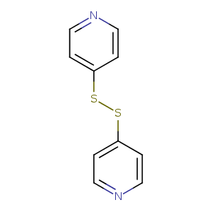
| HET Code: | D4G |
|---|---|
| Formula: | C10H8N2S2 |
| Molecular weight: | 220.314 g/mol |
| DrugBank ID: | DB07623 |
| Buried Surface Area: | 79 % |
| Polar Surface area: | 76.38 Å2 |
| Number of | |
|---|---|
| H-Bond Acceptors: | 4 |
| H-Bond Donors: | 0 |
| Rings: | 2 |
| Aromatic rings: | 2 |
| Anionic atoms: | 0 |
| Cationic atoms: | 0 |
| Rule of Five Violation: | 0 |
| Rotatable Bonds: | 3 |
| X | Y | Z |
|---|---|---|
| 55.4048 | 78.5459 | 59.7905 |
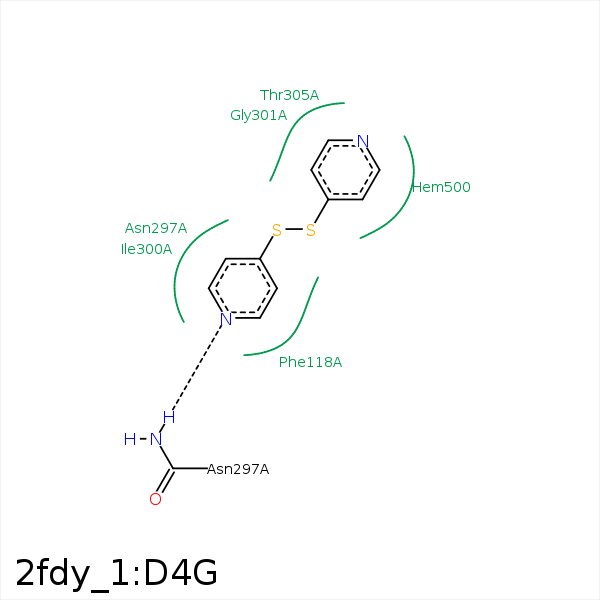


Represent the protein/ligand binding mode, centered on the ligand
Dashed lines represents hydrogen bonds and metal interactions
Green residue labels for amino acids with hydrophobic contacts (green lines) to the ligand
| Ligand | Protein | Interaction | |||
|---|---|---|---|---|---|
| Atom | Atom | Residue | Distance (Å) | Angle (°) | Type |
| S2 | CZ | PHE- 107 | 3.91 | 0 | Hydrophobic |
| C10 | CE2 | PHE- 107 | 3.42 | 0 | Hydrophobic |
| C7 | CG2 | VAL- 117 | 4.16 | 0 | Hydrophobic |
| S1 | CE1 | PHE- 209 | 3.96 | 0 | Hydrophobic |
| N2 | ND2 | ASN- 297 | 2.78 | 140.63 | H-Bond (Protein Donor) |
| S1 | CG2 | ILE- 300 | 4.47 | 0 | Hydrophobic |
| C10 | CG2 | ILE- 300 | 3.89 | 0 | Hydrophobic |
| C5 | CG2 | THR- 305 | 3.36 | 0 | Hydrophobic |
| C3 | CD2 | LEU- 370 | 3.76 | 0 | Hydrophobic |
| S1 | CE2 | PHE- 480 | 4.03 | 0 | Hydrophobic |
| S2 | CZ | PHE- 480 | 3.63 | 0 | Hydrophobic |
