sc-PDB
An Annotated Database of Druggable Binding Sites from the Protein DataBank
An Annotated Database of Druggable Binding Sites from the Protein DataBank
1.990 Å
X-ray
2006-05-10
| Name: | Isocitrate dehydrogenase [NADP] cytoplasmic |
|---|---|
| ID: | IDHC_MOUSE |
| AC: | O88844 |
| Organism: | Mus musculus |
| Reign: | Eukaryota |
| TaxID: | 10090 |
| EC Number: | 1.1.1.42 |
| Chain Name: | Percentage of Residues within binding site |
|---|---|
| A | 81 % |
| B | 19 % |
| B-Factor: | 19.779 |
|---|---|
| Number of residues: | 64 |
| Including | |
| Standard Amino Acids: | 53 |
| Non Standard Amino Acids: | 0 |
| Water Molecules: | 11 |
| Cofactors: | |
| Metals: | |
| Ligandability | Volume (Å3) |
|---|---|
| 0.526 | 1906.875 |
| % Hydrophobic | % Polar |
|---|---|
| 39.12 | 60.88 |
| According to VolSite | |
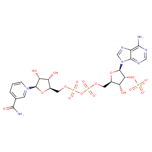
| HET Code: | NAP |
|---|---|
| Formula: | C21H25N7O17P3 |
| Molecular weight: | 740.381 g/mol |
| DrugBank ID: | DB03461 |
| Buried Surface Area: | 62.25 % |
| Polar Surface area: | 405.54 Å2 |
| Number of | |
|---|---|
| H-Bond Acceptors: | 21 |
| H-Bond Donors: | 5 |
| Rings: | 5 |
| Aromatic rings: | 3 |
| Anionic atoms: | 4 |
| Cationic atoms: | 1 |
| Rule of Five Violation: | 2 |
| Rotatable Bonds: | 13 |
| X | Y | Z |
|---|---|---|
| -14.4827 | 8.05181 | 28.3587 |


Represent the protein/ligand binding mode, centered on the ligand
Dashed lines represents hydrogen bonds and metal interactions
Green residue labels for amino acids with hydrophobic contacts (green lines) to the ligand
| Ligand | Protein | Interaction | |||
|---|---|---|---|---|---|
| Atom | Atom | Residue | Distance (Å) | Angle (°) | Type |
| O7N | OG1 | THR- 75 | 3.24 | 158.61 | H-Bond (Protein Donor) |
| O7N | N | THR- 75 | 2.9 | 165.97 | H-Bond (Protein Donor) |
| O2D | N | THR- 77 | 3.22 | 163.23 | H-Bond (Protein Donor) |
| O3D | NH2 | ARG- 82 | 2.88 | 148.28 | H-Bond (Protein Donor) |
| O7N | ND2 | ASN- 96 | 2.86 | 153.84 | H-Bond (Protein Donor) |
| C2D | CD1 | LEU- 250 | 4.25 | 0 | Hydrophobic |
| C5B | CB | ASP- 253 | 4.15 | 0 | Hydrophobic |
| O2B | NE2 | GLN- 257 | 3.4 | 127.79 | H-Bond (Protein Donor) |
| O1X | NE2 | GLN- 257 | 3.27 | 162.03 | H-Bond (Protein Donor) |
| O1X | NZ | LYS- 260 | 2.7 | 164.71 | H-Bond (Protein Donor) |
| O1X | NZ | LYS- 260 | 2.7 | 0 | Ionic (Protein Cationic) |
| C1B | CD1 | LEU- 288 | 4.38 | 0 | Hydrophobic |
| O1A | N | GLY- 310 | 2.79 | 159.88 | H-Bond (Protein Donor) |
| C4D | CB | THR- 311 | 4.18 | 0 | Hydrophobic |
| O4D | N | THR- 311 | 3.09 | 171.4 | H-Bond (Protein Donor) |
| O2A | N | VAL- 312 | 2.78 | 148.12 | H-Bond (Protein Donor) |
| C3B | CG1 | VAL- 312 | 3.85 | 0 | Hydrophobic |
| C5D | CB | THR- 313 | 4.08 | 0 | Hydrophobic |
| O2X | NE2 | HIS- 315 | 2.86 | 173.24 | H-Bond (Protein Donor) |
| N6A | O | ASN- 328 | 2.86 | 162.44 | H-Bond (Ligand Donor) |
| N1A | N | ASN- 328 | 3.06 | 150.76 | H-Bond (Protein Donor) |
| O3D | O | HOH- 2064 | 2.61 | 156.98 | H-Bond (Ligand Donor) |
| O4B | O | HOH- 2167 | 2.74 | 179.98 | H-Bond (Protein Donor) |
| N7N | O | HOH- 2250 | 3.17 | 161.51 | H-Bond (Ligand Donor) |
| O2N | O | HOH- 2253 | 2.68 | 158.12 | H-Bond (Protein Donor) |
| O3X | O | HOH- 2333 | 2.78 | 179.97 | H-Bond (Protein Donor) |
| O1N | O | HOH- 2335 | 2.6 | 179.97 | H-Bond (Protein Donor) |
