sc-PDB
An Annotated Database of Druggable Binding Sites from the Protein DataBank
An Annotated Database of Druggable Binding Sites from the Protein DataBank
3.000 Å
X-ray
2006-04-28
| Name: | Aflatoxin B1 aldehyde reductase member 3 |
|---|---|
| ID: | ARK73_HUMAN |
| AC: | O95154 |
| Organism: | Homo sapiens |
| Reign: | Eukaryota |
| TaxID: | 9606 |
| EC Number: | / |
| Chain Name: | Percentage of Residues within binding site |
|---|---|
| F | 100 % |
| B-Factor: | 55.811 |
|---|---|
| Number of residues: | 51 |
| Including | |
| Standard Amino Acids: | 51 |
| Non Standard Amino Acids: | 0 |
| Water Molecules: | 0 |
| Cofactors: | |
| Metals: | |
| Ligandability | Volume (Å3) |
|---|---|
| 1.143 | 1343.250 |
| % Hydrophobic | % Polar |
|---|---|
| 50.75 | 49.25 |
| According to VolSite | |

| HET Code: | NDP |
|---|---|
| Formula: | C21H26N7O17P3 |
| Molecular weight: | 741.389 g/mol |
| DrugBank ID: | DB02338 |
| Buried Surface Area: | 78.45 % |
| Polar Surface area: | 404.9 Å2 |
| Number of | |
|---|---|
| H-Bond Acceptors: | 22 |
| H-Bond Donors: | 5 |
| Rings: | 5 |
| Aromatic rings: | 2 |
| Anionic atoms: | 4 |
| Cationic atoms: | 0 |
| Rule of Five Violation: | 2 |
| Rotatable Bonds: | 13 |
| X | Y | Z |
|---|---|---|
| -4.68 | 43.4531 | 3.96375 |
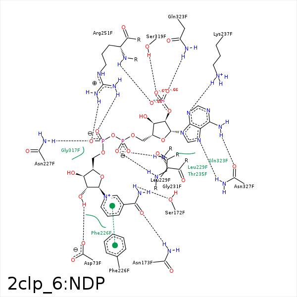


Represent the protein/ligand binding mode, centered on the ligand
Dashed lines represents hydrogen bonds and metal interactions
Green residue labels for amino acids with hydrophobic contacts (green lines) to the ligand
| Ligand | Protein | Interaction | |||
|---|---|---|---|---|---|
| Atom | Atom | Residue | Distance (Å) | Angle (°) | Type |
| O3D | N | MET- 46 | 3.4 | 147.56 | H-Bond (Protein Donor) |
| C5N | SD | MET- 46 | 3.88 | 0 | Hydrophobic |
| C3D | CB | MET- 46 | 3.66 | 0 | Hydrophobic |
| O2X | NE | ARG- 51 | 3.43 | 121.65 | H-Bond (Protein Donor) |
| O2D | OD2 | ASP- 73 | 2.55 | 161.07 | H-Bond (Ligand Donor) |
| C2D | CE1 | TYR- 78 | 4.12 | 0 | Hydrophobic |
| N7N | OG | SER- 172 | 2.7 | 145.83 | H-Bond (Ligand Donor) |
| O7N | ND2 | ASN- 173 | 3.21 | 161.93 | H-Bond (Protein Donor) |
| N7N | OE1 | GLN- 198 | 3.26 | 141.09 | H-Bond (Ligand Donor) |
| C3N | CB | PHE- 226 | 4.26 | 0 | Hydrophobic |
| C5D | CB | PHE- 226 | 4.33 | 0 | Hydrophobic |
| DuAr | DuAr | PHE- 226 | 3.99 | 0 | Aromatic Face/Face |
| O2N | ND2 | ASN- 227 | 2.66 | 120.6 | H-Bond (Protein Donor) |
| O5D | N | ASN- 227 | 3.39 | 160.81 | H-Bond (Protein Donor) |
| O1A | N | LEU- 229 | 2.88 | 144.5 | H-Bond (Protein Donor) |
| O2A | N | LEU- 229 | 3.47 | 133.35 | H-Bond (Protein Donor) |
| O1A | N | GLY- 231 | 2.92 | 148.94 | H-Bond (Protein Donor) |
| N3A | NZ | LYS- 237 | 3.17 | 160.3 | H-Bond (Protein Donor) |
| O3X | NZ | LYS- 237 | 3.45 | 140.1 | H-Bond (Protein Donor) |
| O1X | NZ | LYS- 237 | 3.16 | 0 | Ionic (Protein Cationic) |
| O3X | NZ | LYS- 237 | 3.45 | 0 | Ionic (Protein Cationic) |
| O1N | NH2 | ARG- 251 | 3.14 | 177.55 | H-Bond (Protein Donor) |
| O2N | NH1 | ARG- 251 | 2.97 | 166.96 | H-Bond (Protein Donor) |
| O3X | N | ARG- 251 | 3.12 | 157.91 | H-Bond (Protein Donor) |
| O2N | CZ | ARG- 251 | 3.86 | 0 | Ionic (Protein Cationic) |
| C4D | CG2 | ILE- 315 | 3.55 | 0 | Hydrophobic |
| O2X | OG | SER- 319 | 2.66 | 155.41 | H-Bond (Protein Donor) |
| C3B | CB | SER- 319 | 4.35 | 0 | Hydrophobic |
| N6A | O | GLN- 323 | 3.44 | 130.25 | H-Bond (Ligand Donor) |
| O1X | NE2 | GLN- 323 | 3.02 | 161.23 | H-Bond (Protein Donor) |
| N7A | ND2 | ASN- 327 | 2.72 | 155.52 | H-Bond (Protein Donor) |
| N6A | OD1 | ASN- 327 | 3.15 | 132.38 | H-Bond (Ligand Donor) |
