sc-PDB
An Annotated Database of Druggable Binding Sites from the Protein DataBank
An Annotated Database of Druggable Binding Sites from the Protein DataBank
1.900 Å
X-ray
2005-12-09
| Name: | 6,7-dimethyl-8-ribityllumazine synthase |
|---|---|
| ID: | RISB_MYCTU |
| AC: | P9WHE9 |
| Organism: | Mycobacterium tuberculosis |
| Reign: | Bacteria |
| TaxID: | 83332 |
| EC Number: | 2.5.1.78 |
| Chain Name: | Percentage of Residues within binding site |
|---|---|
| A | 36 % |
| E | 64 % |
| B-Factor: | 30.017 |
|---|---|
| Number of residues: | 41 |
| Including | |
| Standard Amino Acids: | 39 |
| Non Standard Amino Acids: | 0 |
| Water Molecules: | 2 |
| Cofactors: | |
| Metals: | |
| Ligandability | Volume (Å3) |
|---|---|
| 0.732 | 637.875 |
| % Hydrophobic | % Polar |
|---|---|
| 44.97 | 55.03 |
| According to VolSite | |
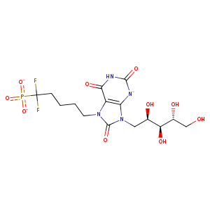
| HET Code: | TSF |
|---|---|
| Formula: | C15H21F2N4O10P |
| Molecular weight: | 486.319 g/mol |
| DrugBank ID: | - |
| Buried Surface Area: | 71.14 % |
| Polar Surface area: | 235.67 Å2 |
| Number of | |
|---|---|
| H-Bond Acceptors: | 10 |
| H-Bond Donors: | 6 |
| Rings: | 2 |
| Aromatic rings: | 0 |
| Anionic atoms: | 2 |
| Cationic atoms: | 0 |
| Rule of Five Violation: | 2 |
| Rotatable Bonds: | 11 |
| X | Y | Z |
|---|---|---|
| 52.9215 | -17.0512 | 11.0928 |
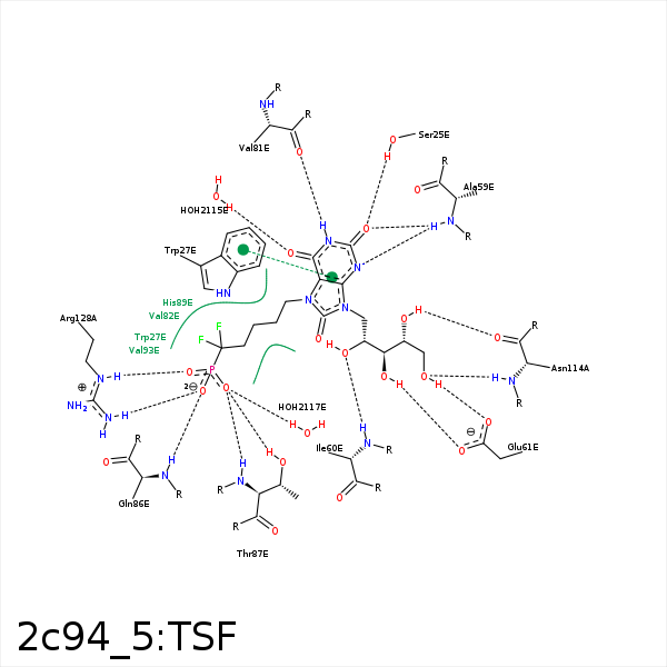


Represent the protein/ligand binding mode, centered on the ligand
Dashed lines represents hydrogen bonds and metal interactions
Green residue labels for amino acids with hydrophobic contacts (green lines) to the ligand
| Ligand | Protein | Interaction | |||
|---|---|---|---|---|---|
| Atom | Atom | Residue | Distance (Å) | Angle (°) | Type |
| O1 | OG | SER- 25 | 3.09 | 150.83 | H-Bond (Protein Donor) |
| C15 | CZ3 | TRP- 27 | 3.48 | 0 | Hydrophobic |
| C10 | CE2 | TRP- 27 | 4.48 | 0 | Hydrophobic |
| O1 | N | ALA- 59 | 2.89 | 137.02 | H-Bond (Protein Donor) |
| O19 | N | ILE- 60 | 3.13 | 174.27 | H-Bond (Protein Donor) |
| C13 | CG1 | ILE- 60 | 4.42 | 0 | Hydrophobic |
| C12 | CG1 | ILE- 60 | 3.81 | 0 | Hydrophobic |
| O26 | OE2 | GLU- 61 | 2.7 | 163.26 | H-Bond (Ligand Donor) |
| N3 | O | VAL- 81 | 2.73 | 174.4 | H-Bond (Ligand Donor) |
| C16 | CG1 | VAL- 82 | 4.36 | 0 | Hydrophobic |
| O2P | N | GLN- 86 | 3 | 171.67 | H-Bond (Protein Donor) |
| O3P | OG1 | THR- 87 | 2.61 | 171.53 | H-Bond (Protein Donor) |
| O3P | N | THR- 87 | 2.78 | 155.93 | H-Bond (Protein Donor) |
| O1P | OG1 | THR- 87 | 3.5 | 125.79 | H-Bond (Protein Donor) |
| C18 | CB | HIS- 89 | 3.85 | 0 | Hydrophobic |
| C17 | CB | PHE- 90 | 4.34 | 0 | Hydrophobic |
| C10 | CG2 | VAL- 93 | 4.43 | 0 | Hydrophobic |
| C14 | CB | ALA- 113 | 4.1 | 0 | Hydrophobic |
| O23 | O | ASN- 114 | 2.81 | 161.19 | H-Bond (Ligand Donor) |
| O26 | N | ASN- 114 | 2.74 | 157.95 | H-Bond (Protein Donor) |
| O2P | NH2 | ARG- 128 | 3.22 | 153.95 | H-Bond (Protein Donor) |
| O1P | NE | ARG- 128 | 3.1 | 160.5 | H-Bond (Protein Donor) |
| O1P | NH2 | ARG- 128 | 3.46 | 138.82 | H-Bond (Protein Donor) |
| O1P | CZ | ARG- 128 | 3.73 | 0 | Ionic (Protein Cationic) |
| C14 | CB | ALA- 142 | 4.23 | 0 | Hydrophobic |
| C14 | CB | ALA- 145 | 4.07 | 0 | Hydrophobic |
| O2 | O | HOH- 2115 | 2.64 | 174.93 | H-Bond (Protein Donor) |
| O3P | O | HOH- 2117 | 2.53 | 179.99 | H-Bond (Protein Donor) |
