sc-PDB
An Annotated Database of Druggable Binding Sites from the Protein DataBank
An Annotated Database of Druggable Binding Sites from the Protein DataBank
2.070 Å
X-ray
2005-09-10
| Name: | Nitroalkane oxidase |
|---|---|
| ID: | NAO_FUSOX |
| AC: | Q8X1D8 |
| Organism: | Fusarium oxysporum |
| Reign: | Eukaryota |
| TaxID: | 5507 |
| EC Number: | 1.7.3.1 |
| Chain Name: | Percentage of Residues within binding site |
|---|---|
| A | 28 % |
| B | 70 % |
| D | 2 % |
| B-Factor: | 21.122 |
|---|---|
| Number of residues: | 67 |
| Including | |
| Standard Amino Acids: | 61 |
| Non Standard Amino Acids: | 0 |
| Water Molecules: | 6 |
| Cofactors: | |
| Metals: | |
| Ligandability | Volume (Å3) |
|---|---|
| 1.404 | 985.500 |
| % Hydrophobic | % Polar |
|---|---|
| 52.74 | 47.26 |
| According to VolSite | |
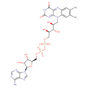
| HET Code: | FAD |
|---|---|
| Formula: | C27H31N9O15P2 |
| Molecular weight: | 783.534 g/mol |
| DrugBank ID: | DB03147 |
| Buried Surface Area: | 70.76 % |
| Polar Surface area: | 381.7 Å2 |
| Number of | |
|---|---|
| H-Bond Acceptors: | 22 |
| H-Bond Donors: | 7 |
| Rings: | 6 |
| Aromatic rings: | 3 |
| Anionic atoms: | 2 |
| Cationic atoms: | 0 |
| Rule of Five Violation: | 3 |
| Rotatable Bonds: | 13 |
| X | Y | Z |
|---|---|---|
| 91.6529 | 28.8753 | 210.455 |


Represent the protein/ligand binding mode, centered on the ligand
Dashed lines represents hydrogen bonds and metal interactions
Green residue labels for amino acids with hydrophobic contacts (green lines) to the ligand
| Ligand | Protein | Interaction | |||
|---|---|---|---|---|---|
| Atom | Atom | Residue | Distance (Å) | Angle (°) | Type |
| N3 | O | LEU- 131 | 2.62 | 153 | H-Bond (Ligand Donor) |
| O2 | N | HIS- 133 | 2.92 | 142.67 | H-Bond (Protein Donor) |
| N1 | OG | SER- 134 | 2.88 | 165.2 | H-Bond (Protein Donor) |
| O2 | N | SER- 134 | 2.73 | 165.94 | H-Bond (Protein Donor) |
| C1' | CB | SER- 134 | 3.99 | 0 | Hydrophobic |
| C3' | CB | SER- 134 | 4.25 | 0 | Hydrophobic |
| O3' | OG | SER- 134 | 2.86 | 134.23 | H-Bond (Ligand Donor) |
| O1A | N | THR- 139 | 3.01 | 134.88 | H-Bond (Protein Donor) |
| O1A | N | ALA- 140 | 3.01 | 162.45 | H-Bond (Protein Donor) |
| N7A | ND2 | ASN- 141 | 3.15 | 139.74 | H-Bond (Protein Donor) |
| N6A | OD1 | ASN- 141 | 2.73 | 166.93 | H-Bond (Ligand Donor) |
| C8M | CE3 | TRP- 169 | 4 | 0 | Hydrophobic |
| C1' | CB | TRP- 169 | 3.38 | 0 | Hydrophobic |
| C9 | CB | TRP- 169 | 3.86 | 0 | Hydrophobic |
| O4 | N | SER- 171 | 3.39 | 152.72 | H-Bond (Protein Donor) |
| N5 | OG | SER- 171 | 2.87 | 139.02 | H-Bond (Protein Donor) |
| C6 | CB | SER- 171 | 4.35 | 0 | Hydrophobic |
| C7M | CD1 | LEU- 234 | 4.18 | 0 | Hydrophobic |
| C8M | CD1 | LEU- 234 | 4.16 | 0 | Hydrophobic |
| C7M | CG2 | THR- 240 | 3.87 | 0 | Hydrophobic |
| O2A | NH1 | ARG- 304 | 3.07 | 156.91 | H-Bond (Protein Donor) |
| C5B | CG | ARG- 304 | 3.32 | 0 | Hydrophobic |
| C1B | CG1 | ILE- 310 | 3.5 | 0 | Hydrophobic |
| DuAr | DuAr | HIS- 313 | 3.76 | 0 | Aromatic Face/Face |
| N1A | NE2 | GLN- 314 | 2.94 | 138.67 | H-Bond (Protein Donor) |
| C1B | CG2 | VAL- 316 | 3.66 | 0 | Hydrophobic |
| O3B | O | LYS- 375 | 3.09 | 136.77 | H-Bond (Ligand Donor) |
| O2B | O | ALA- 376 | 2.73 | 163.25 | H-Bond (Ligand Donor) |
| C8M | SD | MET- 379 | 3.71 | 0 | Hydrophobic |
| C9 | CE | MET- 379 | 3.24 | 0 | Hydrophobic |
| C2' | CE | MET- 379 | 3.27 | 0 | Hydrophobic |
| O2P | N | MET- 379 | 2.91 | 159.94 | H-Bond (Protein Donor) |
| C8M | CD2 | TYR- 382 | 4.3 | 0 | Hydrophobic |
| C7M | SG | CYS- 397 | 4.18 | 0 | Hydrophobic |
| C8M | SG | CYS- 397 | 4.11 | 0 | Hydrophobic |
| O4' | O | LEU- 400 | 2.91 | 172.9 | H-Bond (Ligand Donor) |
| C7M | CD1 | PHE- 401 | 4.37 | 0 | Hydrophobic |
| C8 | CB | PHE- 401 | 4.14 | 0 | Hydrophobic |
| O4' | N | GLY- 403 | 2.92 | 122.71 | H-Bond (Protein Donor) |
| O1P | N | GLY- 404 | 2.79 | 138.71 | H-Bond (Protein Donor) |
| C2B | CG2 | ILE- 406 | 3.91 | 0 | Hydrophobic |
| C5' | CD1 | LEU- 408 | 4.04 | 0 | Hydrophobic |
| O2A | O | HOH- 2195 | 2.77 | 179.96 | H-Bond (Protein Donor) |
| O4 | O | HOH- 2198 | 2.79 | 155.7 | H-Bond (Protein Donor) |
