sc-PDB
An Annotated Database of Druggable Binding Sites from the Protein DataBank
An Annotated Database of Druggable Binding Sites from the Protein DataBank
2.700 Å
X-ray
2005-09-12
| Name: | 2,5-diamino-6-ribosylamino-4(3H)-pyrimidinone 5'-phosphate reductase |
|---|---|
| ID: | RIB7_METJA |
| AC: | Q58085 |
| Organism: | Methanocaldococcus jannaschii |
| Reign: | Archaea |
| TaxID: | 243232 |
| EC Number: | / |
| Chain Name: | Percentage of Residues within binding site |
|---|---|
| A | 100 % |
| B-Factor: | 48.685 |
|---|---|
| Number of residues: | 45 |
| Including | |
| Standard Amino Acids: | 45 |
| Non Standard Amino Acids: | 0 |
| Water Molecules: | 0 |
| Cofactors: | |
| Metals: | |
| Ligandability | Volume (Å3) |
|---|---|
| 0.288 | 418.500 |
| % Hydrophobic | % Polar |
|---|---|
| 36.29 | 63.71 |
| According to VolSite | |
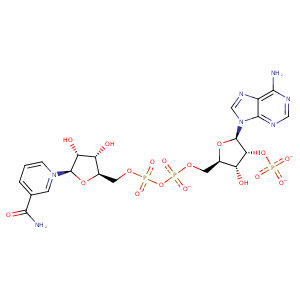
| HET Code: | NAP |
|---|---|
| Formula: | C21H25N7O17P3 |
| Molecular weight: | 740.381 g/mol |
| DrugBank ID: | DB03461 |
| Buried Surface Area: | 63.92 % |
| Polar Surface area: | 405.54 Å2 |
| Number of | |
|---|---|
| H-Bond Acceptors: | 21 |
| H-Bond Donors: | 5 |
| Rings: | 5 |
| Aromatic rings: | 3 |
| Anionic atoms: | 4 |
| Cationic atoms: | 1 |
| Rule of Five Violation: | 2 |
| Rotatable Bonds: | 13 |
| X | Y | Z |
|---|---|---|
| -6.97906 | -51.2956 | -45.2084 |
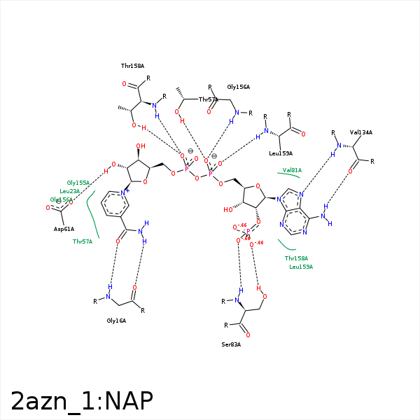


Represent the protein/ligand binding mode, centered on the ligand
Dashed lines represents hydrogen bonds and metal interactions
Green residue labels for amino acids with hydrophobic contacts (green lines) to the ligand
| Ligand | Protein | Interaction | |||
|---|---|---|---|---|---|
| Atom | Atom | Residue | Distance (Å) | Angle (°) | Type |
| C3N | CG2 | VAL- 15 | 4.09 | 0 | Hydrophobic |
| O7N | N | GLY- 16 | 3.15 | 149.49 | H-Bond (Protein Donor) |
| N7N | O | GLY- 16 | 2.84 | 125.86 | H-Bond (Ligand Donor) |
| C3N | CD1 | LEU- 23 | 3.94 | 0 | Hydrophobic |
| O3D | OD1 | ASN- 28 | 3.49 | 143.75 | H-Bond (Ligand Donor) |
| C4B | CG2 | ILE- 55 | 3.62 | 0 | Hydrophobic |
| C1B | CG2 | ILE- 55 | 3.88 | 0 | Hydrophobic |
| O1A | OG1 | THR- 57 | 3.24 | 153.06 | H-Bond (Protein Donor) |
| O1A | N | THR- 57 | 3.49 | 141.43 | H-Bond (Protein Donor) |
| C3D | CB | THR- 57 | 4.45 | 0 | Hydrophobic |
| C2D | CG2 | THR- 57 | 4.25 | 0 | Hydrophobic |
| C5N | CG2 | THR- 57 | 3.96 | 0 | Hydrophobic |
| O2N | NZ | LYS- 60 | 3.96 | 0 | Ionic (Protein Cationic) |
| C3D | CD | LYS- 60 | 4.03 | 0 | Hydrophobic |
| O2D | OD1 | ASP- 61 | 2.97 | 164.35 | H-Bond (Ligand Donor) |
| O2X | N | SER- 83 | 2.78 | 138.03 | H-Bond (Protein Donor) |
| O3X | OG | SER- 83 | 3.08 | 138.27 | H-Bond (Protein Donor) |
| O1X | NZ | LYS- 84 | 3.36 | 153.73 | H-Bond (Protein Donor) |
| O1X | NZ | LYS- 84 | 3.36 | 0 | Ionic (Protein Cationic) |
| O3X | NZ | LYS- 84 | 3.57 | 0 | Ionic (Protein Cationic) |
| O1X | CZ | ARG- 86 | 3.93 | 0 | Ionic (Protein Cationic) |
| N7A | N | VAL- 134 | 3.22 | 146.4 | H-Bond (Protein Donor) |
| N6A | O | VAL- 134 | 2.95 | 143.55 | H-Bond (Ligand Donor) |
| O1A | N | GLY- 156 | 2.8 | 140.88 | H-Bond (Protein Donor) |
| C3B | CG2 | THR- 158 | 3.69 | 0 | Hydrophobic |
| O1N | N | THR- 158 | 2.83 | 151.08 | H-Bond (Protein Donor) |
| O1N | OG1 | THR- 158 | 2.65 | 154.46 | H-Bond (Protein Donor) |
| O2A | N | LEU- 159 | 2.75 | 157.22 | H-Bond (Protein Donor) |
| C5D | CB | PRO- 187 | 3.9 | 0 | Hydrophobic |
