sc-PDB
An Annotated Database of Druggable Binding Sites from the Protein DataBank
An Annotated Database of Druggable Binding Sites from the Protein DataBank
2.300 Å
X-ray
2005-08-18
| Name: | Enoyl-[acyl-carrier-protein] reductase [NADH] |
|---|---|
| ID: | INHA_MYCTU |
| AC: | P9WGR1 |
| Organism: | Mycobacterium tuberculosis |
| Reign: | Bacteria |
| TaxID: | 83332 |
| EC Number: | 1.3.1.9 |
| Chain Name: | Percentage of Residues within binding site |
|---|---|
| A | 100 % |
| B-Factor: | 25.895 |
|---|---|
| Number of residues: | 51 |
| Including | |
| Standard Amino Acids: | 48 |
| Non Standard Amino Acids: | 0 |
| Water Molecules: | 3 |
| Cofactors: | |
| Metals: | |
| Ligandability | Volume (Å3) |
|---|---|
| 1.678 | 1009.125 |
| % Hydrophobic | % Polar |
|---|---|
| 59.87 | 40.13 |
| According to VolSite | |
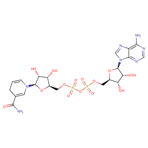
| HET Code: | NAI |
|---|---|
| Formula: | C21H27N7O14P2 |
| Molecular weight: | 663.425 g/mol |
| DrugBank ID: | DB00157 |
| Buried Surface Area: | 68.54 % |
| Polar Surface area: | 342.9 Å2 |
| Number of | |
|---|---|
| H-Bond Acceptors: | 19 |
| H-Bond Donors: | 6 |
| Rings: | 5 |
| Aromatic rings: | 2 |
| Anionic atoms: | 2 |
| Cationic atoms: | 0 |
| Rule of Five Violation: | 3 |
| Rotatable Bonds: | 11 |
| X | Y | Z |
|---|---|---|
| 2.23507 | -32.3913 | 13.939 |
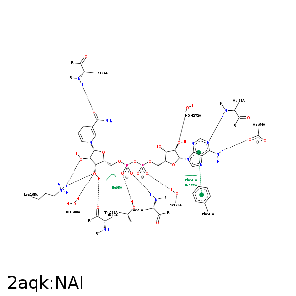


Represent the protein/ligand binding mode, centered on the ligand
Dashed lines represents hydrogen bonds and metal interactions
Green residue labels for amino acids with hydrophobic contacts (green lines) to the ligand
| Ligand | Protein | Interaction | |||
|---|---|---|---|---|---|
| Atom | Atom | Residue | Distance (Å) | Angle (°) | Type |
| C3B | CG2 | ILE- 16 | 3.59 | 0 | Hydrophobic |
| C2B | CB | ILE- 16 | 4.33 | 0 | Hydrophobic |
| O2A | OG | SER- 20 | 2.56 | 165.56 | H-Bond (Protein Donor) |
| O2N | N | ILE- 21 | 2.74 | 168.06 | H-Bond (Protein Donor) |
| C4N | CD1 | ILE- 21 | 4.46 | 0 | Hydrophobic |
| N6A | OD2 | ASP- 64 | 2.95 | 147.04 | H-Bond (Ligand Donor) |
| N1A | N | VAL- 65 | 2.85 | 165.3 | H-Bond (Protein Donor) |
| C5D | CB | ALA- 94 | 3.99 | 0 | Hydrophobic |
| C1B | CG1 | ILE- 95 | 4.07 | 0 | Hydrophobic |
| O3D | O | ILE- 95 | 3.18 | 137.24 | H-Bond (Ligand Donor) |
| O4B | N | GLY- 96 | 3.38 | 156.05 | H-Bond (Protein Donor) |
| C1D | CB | MET- 147 | 3.55 | 0 | Hydrophobic |
| C4D | CB | MET- 147 | 3.45 | 0 | Hydrophobic |
| C4N | CD1 | PHE- 149 | 3.68 | 0 | Hydrophobic |
| O3D | NZ | LYS- 165 | 3.08 | 133.67 | H-Bond (Protein Donor) |
| O2D | NZ | LYS- 165 | 2.87 | 138.38 | H-Bond (Protein Donor) |
| C4N | CB | ALA- 191 | 4.19 | 0 | Hydrophobic |
| O7N | N | ILE- 194 | 2.52 | 168.1 | H-Bond (Protein Donor) |
| N7N | O | ILE- 194 | 3.21 | 138.15 | H-Bond (Ligand Donor) |
| O1N | OG1 | THR- 196 | 3.07 | 161.39 | H-Bond (Protein Donor) |
| O2B | O | HOH- 272 | 2.57 | 158.9 | H-Bond (Protein Donor) |
| O3D | O | HOH- 287 | 3.44 | 128.09 | H-Bond (Ligand Donor) |
| O3D | O | HOH- 288 | 2.8 | 134.87 | H-Bond (Protein Donor) |
