sc-PDB
An Annotated Database of Druggable Binding Sites from the Protein DataBank
An Annotated Database of Druggable Binding Sites from the Protein DataBank
2.600 Å
X-ray
2005-07-01
| Name: | Cell death protein 4 |
|---|---|
| ID: | CED4_CAEEL |
| AC: | P30429 |
| Organism: | Caenorhabditis elegans |
| Reign: | Eukaryota |
| TaxID: | 6239 |
| EC Number: | / |
| Chain Name: | Percentage of Residues within binding site |
|---|---|
| B | 100 % |
| B-Factor: | 75.693 |
|---|---|
| Number of residues: | 40 |
| Including | |
| Standard Amino Acids: | 39 |
| Non Standard Amino Acids: | 1 |
| Water Molecules: | 0 |
| Cofactors: | |
| Metals: | MG |
| Ligandability | Volume (Å3) |
|---|---|
| 0.537 | 1971.000 |
| % Hydrophobic | % Polar |
|---|---|
| 50.68 | 49.32 |
| According to VolSite | |
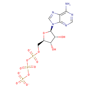
| HET Code: | ATP |
|---|---|
| Formula: | C10H12N5O13P3 |
| Molecular weight: | 503.149 g/mol |
| DrugBank ID: | DB00171 |
| Buried Surface Area: | 74.05 % |
| Polar Surface area: | 319.88 Å2 |
| Number of | |
|---|---|
| H-Bond Acceptors: | 17 |
| H-Bond Donors: | 3 |
| Rings: | 3 |
| Aromatic rings: | 2 |
| Anionic atoms: | 4 |
| Cationic atoms: | 0 |
| Rule of Five Violation: | 2 |
| Rotatable Bonds: | 8 |
| X | Y | Z |
|---|---|---|
| 96.0145 | 48.6215 | 75.4147 |
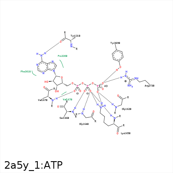


Represent the protein/ligand binding mode, centered on the ligand
Dashed lines represents hydrogen bonds and metal interactions
Green residue labels for amino acids with hydrophobic contacts (green lines) to the ligand
| Ligand | Protein | Interaction | |||
|---|---|---|---|---|---|
| Atom | Atom | Residue | Distance (Å) | Angle (°) | Type |
| N6 | O | TYR- 131 | 2.91 | 160.61 | H-Bond (Ligand Donor) |
| O1G | N | GLY- 162 | 3.28 | 160.37 | H-Bond (Protein Donor) |
| O2B | N | GLY- 162 | 3.27 | 137.15 | H-Bond (Protein Donor) |
| O3B | N | GLY- 162 | 3.26 | 125.79 | H-Bond (Protein Donor) |
| O2B | N | GLY- 164 | 3.19 | 158.78 | H-Bond (Protein Donor) |
| O2B | N | LYS- 165 | 3.21 | 163.68 | H-Bond (Protein Donor) |
| O1B | N | SER- 166 | 3.1 | 165.46 | H-Bond (Protein Donor) |
| O1A | N | VAL- 167 | 3.01 | 161.77 | H-Bond (Protein Donor) |
| C5' | CG2 | VAL- 167 | 3.38 | 0 | Hydrophobic |
| O3G | NH2 | ARG- 273 | 2.71 | 172.37 | H-Bond (Protein Donor) |
| O3G | NE | ARG- 273 | 3.42 | 130.51 | H-Bond (Protein Donor) |
| O3G | CZ | ARG- 273 | 3.49 | 0 | Ionic (Protein Cationic) |
| C1' | CB | PRO- 330 | 4.13 | 0 | Hydrophobic |
| C4' | CB | ALA- 331 | 4.18 | 0 | Hydrophobic |
| C1' | CB | MET- 334 | 4.5 | 0 | Hydrophobic |
| C2' | CG | PRO- 368 | 4.35 | 0 | Hydrophobic |
| O3G | OH | TYR- 369 | 2.62 | 139.91 | H-Bond (Protein Donor) |
| C3' | CB | TYR- 369 | 3.5 | 0 | Hydrophobic |
| C4' | CD1 | TYR- 369 | 3.53 | 0 | Hydrophobic |
| O2G | MG | MG- 550 | 2.3 | 0 | Metal Acceptor |
| O1B | MG | MG- 550 | 2.37 | 0 | Metal Acceptor |
