sc-PDB
An Annotated Database of Druggable Binding Sites from the Protein DataBank
An Annotated Database of Druggable Binding Sites from the Protein DataBank
2.100 Å
X-ray
2005-06-30
| Name: | L-lysine 2,3-aminomutase |
|---|---|
| ID: | KAMA_CLOSU |
| AC: | Q9XBQ8 |
| Organism: | Clostridium subterminale |
| Reign: | Bacteria |
| TaxID: | 1550 |
| EC Number: | 5.4.3.2 |
| Chain Name: | Percentage of Residues within binding site |
|---|---|
| A | 100 % |
| B-Factor: | 29.850 |
|---|---|
| Number of residues: | 43 |
| Including | |
| Standard Amino Acids: | 39 |
| Non Standard Amino Acids: | 1 |
| Water Molecules: | 3 |
| Cofactors: | |
| Metals: | |
| Ligandability | Volume (Å3) |
|---|---|
| 0.300 | 688.500 |
| % Hydrophobic | % Polar |
|---|---|
| 29.41 | 70.59 |
| According to VolSite | |
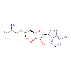
| HET Code: | SAM |
|---|---|
| Formula: | C15H23N6O5S |
| Molecular weight: | 399.445 g/mol |
| DrugBank ID: | DB00118 |
| Buried Surface Area: | 80.61 % |
| Polar Surface area: | 189.77 Å2 |
| Number of | |
|---|---|
| H-Bond Acceptors: | 9 |
| H-Bond Donors: | 4 |
| Rings: | 3 |
| Aromatic rings: | 2 |
| Anionic atoms: | 1 |
| Cationic atoms: | 2 |
| Rule of Five Violation: | 1 |
| Rotatable Bonds: | 7 |
| X | Y | Z |
|---|---|---|
| -30.7661 | 0.745296 | -16.9862 |
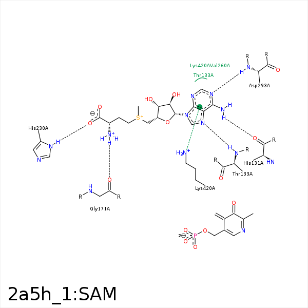


Represent the protein/ligand binding mode, centered on the ligand
Dashed lines represents hydrogen bonds and metal interactions
Green residue labels for amino acids with hydrophobic contacts (green lines) to the ligand
| Ligand | Protein | Interaction | |||
|---|---|---|---|---|---|
| Atom | Atom | Residue | Distance (Å) | Angle (°) | Type |
| N6 | O | HIS- 131 | 2.81 | 164.82 | H-Bond (Ligand Donor) |
| N7 | N | THR- 133 | 3.14 | 145.44 | H-Bond (Protein Donor) |
| CE | CB | ARG- 134 | 4.11 | 0 | Hydrophobic |
| N | O | GLY- 171 | 3.18 | 151.47 | H-Bond (Ligand Donor) |
| OXT | ND1 | HIS- 230 | 2.96 | 153.04 | H-Bond (Protein Donor) |
| C3' | CB | HIS- 230 | 4.14 | 0 | Hydrophobic |
| C1' | CD1 | TYR- 290 | 4.13 | 0 | Hydrophobic |
| N1 | N | ASP- 293 | 2.88 | 152.3 | H-Bond (Protein Donor) |
