sc-PDB
An Annotated Database of Druggable Binding Sites from the Protein DataBank
An Annotated Database of Druggable Binding Sites from the Protein DataBank
2.020 Å
X-ray
2005-05-16
| Name: | Protease |
|---|---|
| ID: | O92139_9HIV1 |
| AC: | O92139 |
| Organism: | Human immunodeficiency virus 1 |
| Reign: | Viruses |
| TaxID: | 11676 |
| EC Number: | / |
| Chain Name: | Percentage of Residues within binding site |
|---|---|
| A | 100 % |
| B-Factor: | 24.333 |
|---|---|
| Number of residues: | 23 |
| Including | |
| Standard Amino Acids: | 23 |
| Non Standard Amino Acids: | 0 |
| Water Molecules: | 0 |
| Cofactors: | |
| Metals: | |
| Ligandability | Volume (Å3) |
|---|---|
| 0.427 | 334.125 |
| % Hydrophobic | % Polar |
|---|---|
| 52.53 | 47.47 |
| According to VolSite | |
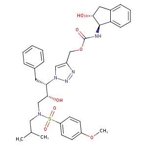
| HET Code: | AB2 |
|---|---|
| Formula: | C34H41N5O7S |
| Molecular weight: | 663.784 g/mol |
| DrugBank ID: | - |
| Buried Surface Area: | 32.73 % |
| Polar Surface area: | 164.48 Å2 |
| Number of | |
|---|---|
| H-Bond Acceptors: | 9 |
| H-Bond Donors: | 3 |
| Rings: | 5 |
| Aromatic rings: | 4 |
| Anionic atoms: | 0 |
| Cationic atoms: | 0 |
| Rule of Five Violation: | 2 |
| Rotatable Bonds: | 15 |
| X | Y | Z |
|---|---|---|
| -10.3066 | 15.0296 | 28.3587 |
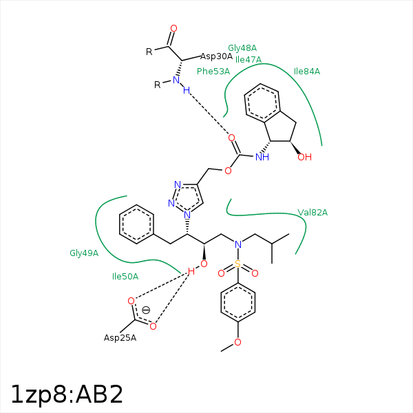


Represent the protein/ligand binding mode, centered on the ligand
Dashed lines represents hydrogen bonds and metal interactions
Green residue labels for amino acids with hydrophobic contacts (green lines) to the ligand
| Ligand | Protein | Interaction | |||
|---|---|---|---|---|---|
| Atom | Atom | Residue | Distance (Å) | Angle (°) | Type |
| O4 | OD1 | ASP- 25 | 2.64 | 132.23 | H-Bond (Ligand Donor) |
| O4 | OD2 | ASP- 25 | 2.74 | 145.35 | H-Bond (Ligand Donor) |
| C24 | CB | ALA- 28 | 4 | 0 | Hydrophobic |
| O6 | N | ASP- 30 | 2.77 | 157.56 | H-Bond (Protein Donor) |
| C24 | CG2 | VAL- 32 | 4.44 | 0 | Hydrophobic |
| C33 | CB | ILE- 47 | 4.21 | 0 | Hydrophobic |
| C17 | CD1 | ILE- 50 | 3.98 | 0 | Hydrophobic |
| C18 | CG1 | ILE- 50 | 3.58 | 0 | Hydrophobic |
| C11 | CG1 | VAL- 82 | 3.29 | 0 | Hydrophobic |
| C11 | CD1 | ILE- 84 | 3.3 | 0 | Hydrophobic |
