
sc-PDB
An Annotated Database of Druggable Binding Sites from the Protein DataBank


An Annotated Database of Druggable Binding Sites from the Protein DataBank
2.300 Å
X-ray
2005-05-12
| Name: | Guanylate kinase |
|---|---|
| ID: | KGUA_MYCTU |
| AC: | P9WKE9 |
| Organism: | Mycobacterium tuberculosis |
| Reign: | Bacteria |
| TaxID: | 83332 |
| EC Number: | 2.7.4.8 |
| Chain Name: | Percentage of Residues within binding site |
|---|---|
| A | 100 % |
| B-Factor: | 24.232 |
|---|---|
| Number of residues: | 22 |
| Including | |
| Standard Amino Acids: | 21 |
| Non Standard Amino Acids: | 0 |
| Water Molecules: | 1 |
| Cofactors: | |
| Metals: | |
| Ligandability | Volume (Å3) |
|---|---|
| 0.046 | 381.375 |
| % Hydrophobic | % Polar |
|---|---|
| 30.09 | 69.91 |
| According to VolSite | |
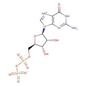
| HET Code: | GDP |
|---|---|
| Formula: | C10H12N5O11P2 |
| Molecular weight: | 440.177 g/mol |
| DrugBank ID: | DB04315 |
| Buried Surface Area: | 37.74 % |
| Polar Surface area: | 276.39 Å2 |
| Number of | |
|---|---|
| H-Bond Acceptors: | 14 |
| H-Bond Donors: | 4 |
| Rings: | 3 |
| Aromatic rings: | 1 |
| Anionic atoms: | 3 |
| Cationic atoms: | 0 |
| Rule of Five Violation: | 1 |
| Rotatable Bonds: | 6 |
| X | Y | Z |
|---|---|---|
| 25.7164 | 8.15311 | 31.7074 |
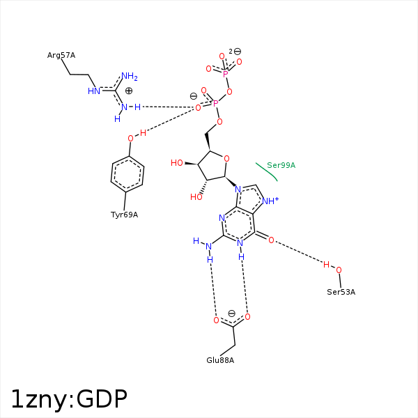
 Image generated by PoseView


Represent the protein/ligand binding mode, centered on the ligand
Dashed lines represents hydrogen bonds and metal interactions
Green residue labels for amino acids with hydrophobic contacts (green lines) to the ligand
| Ligand | Protein | Interaction | |||
|---|---|---|---|---|---|
| Atom | Atom | Residue | Distance (Å) | Angle (°) | Type |
| O6 | OG | SER- 53 | 2.65 | 167.35 | H-Bond (Protein Donor) |
| O1A | NH2 | ARG- 57 | 2.98 | 153.23 | H-Bond (Protein Donor) |
| O2A | NH2 | ARG- 60 | 3.42 | 167.07 | H-Bond (Protein Donor) |
| O1A | OH | TYR- 69 | 3 | 139.24 | H-Bond (Protein Donor) |
| N1 | OE1 | GLU- 88 | 2.72 | 157.58 | H-Bond (Ligand Donor) |
| N1 | OE2 | GLU- 88 | 3.34 | 131.4 | H-Bond (Ligand Donor) |
| N2 | OE2 | GLU- 88 | 2.77 | 155.62 | H-Bond (Ligand Donor) |
| C4' | CG2 | ILE- 92 | 4.07 | 0 | Hydrophobic |
| C1' | CD1 | ILE- 92 | 3.83 | 0 | Hydrophobic |