sc-PDB
An Annotated Database of Druggable Binding Sites from the Protein DataBank
An Annotated Database of Druggable Binding Sites from the Protein DataBank
1.600 Å
X-ray
2005-05-02
| Name: | Mercuric reductase |
|---|---|
| ID: | MERA_PSEAI |
| AC: | P00392 |
| Organism: | Pseudomonas aeruginosa |
| Reign: | Bacteria |
| TaxID: | 287 |
| EC Number: | 1.16.1.1 |
| Chain Name: | Percentage of Residues within binding site |
|---|---|
| A | 100 % |
| B-Factor: | 14.189 |
|---|---|
| Number of residues: | 67 |
| Including | |
| Standard Amino Acids: | 62 |
| Non Standard Amino Acids: | 0 |
| Water Molecules: | 5 |
| Cofactors: | |
| Metals: | |
| Ligandability | Volume (Å3) |
|---|---|
| 0.989 | 1545.750 |
| % Hydrophobic | % Polar |
|---|---|
| 43.01 | 56.99 |
| According to VolSite | |
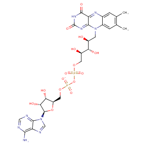
| HET Code: | FAD |
|---|---|
| Formula: | C27H31N9O15P2 |
| Molecular weight: | 783.534 g/mol |
| DrugBank ID: | DB03147 |
| Buried Surface Area: | 73.28 % |
| Polar Surface area: | 381.7 Å2 |
| Number of | |
|---|---|
| H-Bond Acceptors: | 22 |
| H-Bond Donors: | 7 |
| Rings: | 6 |
| Aromatic rings: | 3 |
| Anionic atoms: | 2 |
| Cationic atoms: | 0 |
| Rule of Five Violation: | 3 |
| Rotatable Bonds: | 13 |
| X | Y | Z |
|---|---|---|
| 51.4229 | -18.0079 | 45.185 |
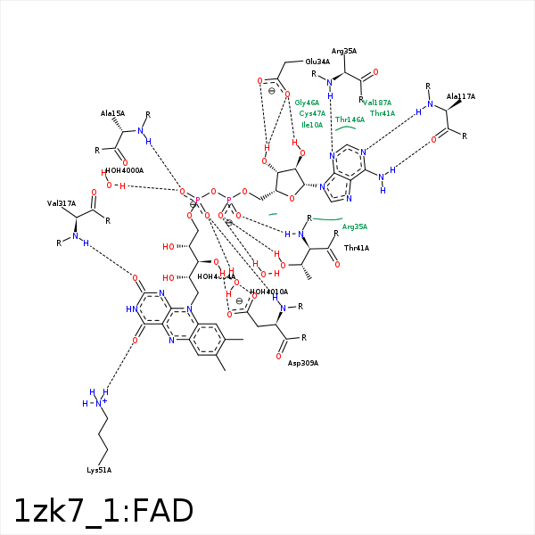


Represent the protein/ligand binding mode, centered on the ligand
Dashed lines represents hydrogen bonds and metal interactions
Green residue labels for amino acids with hydrophobic contacts (green lines) to the ligand
| Ligand | Protein | Interaction | |||
|---|---|---|---|---|---|
| Atom | Atom | Residue | Distance (Å) | Angle (°) | Type |
| O1P | N | ALA- 15 | 2.97 | 165.65 | H-Bond (Protein Donor) |
| O3B | OE2 | GLU- 34 | 2.52 | 158.52 | H-Bond (Ligand Donor) |
| O3B | OE1 | GLU- 34 | 2.98 | 126.48 | H-Bond (Ligand Donor) |
| O2B | OE1 | GLU- 34 | 2.84 | 167.2 | H-Bond (Ligand Donor) |
| C1B | CG | ARG- 35 | 4.45 | 0 | Hydrophobic |
| N3A | N | ARG- 35 | 3.22 | 145.52 | H-Bond (Protein Donor) |
| O1A | OG1 | THR- 41 | 2.66 | 158.99 | H-Bond (Protein Donor) |
| O2A | N | THR- 41 | 2.89 | 166.37 | H-Bond (Protein Donor) |
| C8M | CG2 | THR- 41 | 3.71 | 0 | Hydrophobic |
| O4' | N | CYS- 42 | 3.31 | 136.57 | H-Bond (Protein Donor) |
| C9A | SG | CYS- 47 | 4.45 | 0 | Hydrophobic |
| C2' | SG | CYS- 47 | 4.04 | 0 | Hydrophobic |
| C6 | CB | SER- 50 | 4.4 | 0 | Hydrophobic |
| O4 | NZ | LYS- 51 | 3 | 177.38 | H-Bond (Protein Donor) |
| N6A | O | ALA- 117 | 3.03 | 155.73 | H-Bond (Ligand Donor) |
| N1A | N | ALA- 117 | 3.02 | 168.52 | H-Bond (Protein Donor) |
| C7M | CB | SER- 166 | 3.85 | 0 | Hydrophobic |
| C7M | CD1 | LEU- 170 | 4.16 | 0 | Hydrophobic |
| C7M | CG1 | VAL- 187 | 3.94 | 0 | Hydrophobic |
| C7 | CG2 | VAL- 187 | 3.85 | 0 | Hydrophobic |
| O3' | OD2 | ASP- 309 | 2.94 | 150.02 | H-Bond (Ligand Donor) |
| O3' | OD1 | ASP- 309 | 2.93 | 144.92 | H-Bond (Ligand Donor) |
| C5' | CB | ASP- 309 | 4.36 | 0 | Hydrophobic |
| O2P | N | ASP- 309 | 3.01 | 155.21 | H-Bond (Protein Donor) |
| N1 | N | VAL- 317 | 3.39 | 149.21 | H-Bond (Protein Donor) |
| O2 | N | VAL- 317 | 2.83 | 143.54 | H-Bond (Protein Donor) |
| C2' | CB | VAL- 317 | 4.34 | 0 | Hydrophobic |
| C4' | CB | VAL- 317 | 4.17 | 0 | Hydrophobic |
| C5' | CB | ALA- 320 | 4.25 | 0 | Hydrophobic |
| O1P | O | HOH- 4000 | 2.74 | 171.5 | H-Bond (Protein Donor) |
| O2P | O | HOH- 4004 | 2.72 | 165.89 | H-Bond (Protein Donor) |
| O1A | O | HOH- 4010 | 2.7 | 168.63 | H-Bond (Protein Donor) |
