sc-PDB
An Annotated Database of Druggable Binding Sites from the Protein DataBank
An Annotated Database of Druggable Binding Sites from the Protein DataBank
1.850 Å
X-ray
2005-02-17
| Name: | Methionine aminopeptidase 2 |
|---|---|
| ID: | MAP2_HUMAN |
| AC: | P50579 |
| Organism: | Homo sapiens |
| Reign: | Eukaryota |
| TaxID: | 9606 |
| EC Number: | / |
| Chain Name: | Percentage of Residues within binding site |
|---|---|
| A | 100 % |
| B-Factor: | 19.517 |
|---|---|
| Number of residues: | 30 |
| Including | |
| Standard Amino Acids: | 27 |
| Non Standard Amino Acids: | 2 |
| Water Molecules: | 1 |
| Cofactors: | |
| Metals: | MN MN |
| Ligandability | Volume (Å3) |
|---|---|
| 1.005 | 438.750 |
| % Hydrophobic | % Polar |
|---|---|
| 59.23 | 40.77 |
| According to VolSite | |
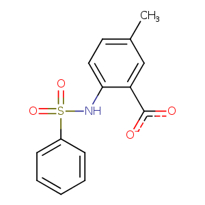
| HET Code: | A41 |
|---|---|
| Formula: | C14H12NO4S |
| Molecular weight: | 290.314 g/mol |
| DrugBank ID: | DB07313 |
| Buried Surface Area: | 64.42 % |
| Polar Surface area: | 94.68 Å2 |
| Number of | |
|---|---|
| H-Bond Acceptors: | 4 |
| H-Bond Donors: | 1 |
| Rings: | 2 |
| Aromatic rings: | 2 |
| Anionic atoms: | 1 |
| Cationic atoms: | 0 |
| Rule of Five Violation: | 0 |
| Rotatable Bonds: | 4 |
| X | Y | Z |
|---|---|---|
| 16.9036 | 29.6255 | 18.206 |
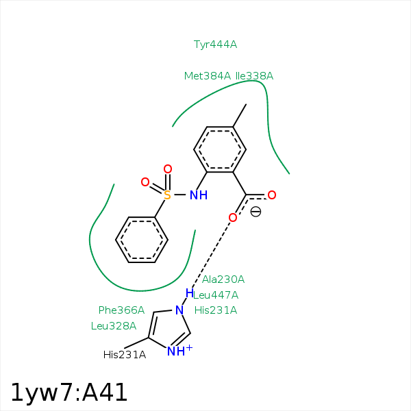


Represent the protein/ligand binding mode, centered on the ligand
Dashed lines represents hydrogen bonds and metal interactions
Green residue labels for amino acids with hydrophobic contacts (green lines) to the ligand
| Ligand | Protein | Interaction | |||
|---|---|---|---|---|---|
| Atom | Atom | Residue | Distance (Å) | Angle (°) | Type |
| C1 | CB | ALA- 230 | 4.01 | 0 | Hydrophobic |
| O18 | NE2 | HIS- 231 | 2.9 | 162.82 | H-Bond (Protein Donor) |
| C1 | CD2 | LEU- 328 | 3.61 | 0 | Hydrophobic |
| C13 | CG2 | ILE- 338 | 3.77 | 0 | Hydrophobic |
| C16 | CG1 | ILE- 338 | 4.02 | 0 | Hydrophobic |
| C12 | CB | HIS- 339 | 4.04 | 0 | Hydrophobic |
| C13 | CE | MET- 384 | 3.79 | 0 | Hydrophobic |
| C20 | CB | ALA- 414 | 3.89 | 0 | Hydrophobic |
| C20 | CG | TYR- 444 | 3.85 | 0 | Hydrophobic |
| C2 | CD1 | LEU- 447 | 3.7 | 0 | Hydrophobic |
| O19 | MN | MN- 481 | 2.35 | 0 | Metal Acceptor |
