sc-PDB
An Annotated Database of Druggable Binding Sites from the Protein DataBank
An Annotated Database of Druggable Binding Sites from the Protein DataBank
2.400 Å
X-ray
2004-12-28
| Name: | NAD(P)H-dependent D-xylose reductase |
|---|---|
| ID: | XYL1_CANTE |
| AC: | O74237 |
| Organism: | Candida tenuis |
| Reign: | Eukaryota |
| TaxID: | 45596 |
| EC Number: | 1.1.1.307 |
| Chain Name: | Percentage of Residues within binding site |
|---|---|
| A | 100 % |
| B-Factor: | 31.237 |
|---|---|
| Number of residues: | 46 |
| Including | |
| Standard Amino Acids: | 44 |
| Non Standard Amino Acids: | 0 |
| Water Molecules: | 2 |
| Cofactors: | |
| Metals: | |
| Ligandability | Volume (Å3) |
|---|---|
| 0.565 | 907.875 |
| % Hydrophobic | % Polar |
|---|---|
| 44.98 | 55.02 |
| According to VolSite | |
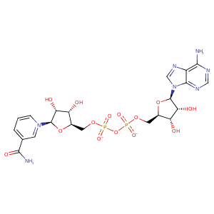
| HET Code: | NAD |
|---|---|
| Formula: | C21H26N7O14P2 |
| Molecular weight: | 662.417 g/mol |
| DrugBank ID: | - |
| Buried Surface Area: | 72.83 % |
| Polar Surface area: | 343.54 Å2 |
| Number of | |
|---|---|
| H-Bond Acceptors: | 18 |
| H-Bond Donors: | 6 |
| Rings: | 5 |
| Aromatic rings: | 3 |
| Anionic atoms: | 2 |
| Cationic atoms: | 1 |
| Rule of Five Violation: | 3 |
| Rotatable Bonds: | 11 |
| X | Y | Z |
|---|---|---|
| 115.559 | 11.2921 | 6.98995 |


Represent the protein/ligand binding mode, centered on the ligand
Dashed lines represents hydrogen bonds and metal interactions
Green residue labels for amino acids with hydrophobic contacts (green lines) to the ligand
| Ligand | Protein | Interaction | |||
|---|---|---|---|---|---|
| Atom | Atom | Residue | Distance (Å) | Angle (°) | Type |
| O2D | N | CYS- 23 | 3.28 | 144.25 | H-Bond (Protein Donor) |
| O3D | N | TRP- 24 | 3.24 | 150.54 | H-Bond (Protein Donor) |
| C2D | CB | TRP- 24 | 3.61 | 0 | Hydrophobic |
| O2D | OD2 | ASP- 47 | 2.87 | 171.81 | H-Bond (Ligand Donor) |
| C2D | CZ | TYR- 52 | 4.28 | 0 | Hydrophobic |
| N7N | OG | SER- 169 | 3.11 | 140.53 | H-Bond (Ligand Donor) |
| O7N | ND2 | ASN- 170 | 3.03 | 174.12 | H-Bond (Protein Donor) |
| N7N | OE1 | GLN- 191 | 2.85 | 141.3 | H-Bond (Ligand Donor) |
| C3N | CB | TYR- 217 | 4.27 | 0 | Hydrophobic |
| C5N | CB | TYR- 217 | 4.38 | 0 | Hydrophobic |
| DuAr | DuAr | TYR- 217 | 3.69 | 0 | Aromatic Face/Face |
| O2N | OG | SER- 218 | 3.46 | 165 | H-Bond (Protein Donor) |
| O5D | N | SER- 218 | 3.33 | 142.7 | H-Bond (Protein Donor) |
| O1A | N | PHE- 220 | 3.46 | 152.5 | H-Bond (Protein Donor) |
| C5B | CD1 | PHE- 220 | 4.14 | 0 | Hydrophobic |
| C1B | CG | GLN- 223 | 4.2 | 0 | Hydrophobic |
| C4B | CG | GLN- 223 | 3.56 | 0 | Hydrophobic |
| C5B | CB | SER- 224 | 3.64 | 0 | Hydrophobic |
| O2N | OG | SER- 224 | 2.56 | 140.16 | H-Bond (Protein Donor) |
| O3B | OE2 | GLU- 227 | 3.1 | 134.76 | H-Bond (Ligand Donor) |
| O3B | OE1 | GLU- 227 | 2.56 | 149.45 | H-Bond (Ligand Donor) |
| O2B | OE2 | GLU- 227 | 2.59 | 166.77 | H-Bond (Ligand Donor) |
| C2D | CD1 | ILE- 272 | 4.33 | 0 | Hydrophobic |
| C4D | CG1 | ILE- 272 | 3.76 | 0 | Hydrophobic |
| O2A | N | ARG- 274 | 2.83 | 150.5 | H-Bond (Protein Donor) |
| O2B | ND2 | ASN- 276 | 3.04 | 178.09 | H-Bond (Protein Donor) |
| O2B | NH1 | ARG- 280 | 3.34 | 121.59 | H-Bond (Protein Donor) |
| DuAr | CZ | ARG- 280 | 3.64 | 153.41 | Pi/Cation |
| N7A | ND2 | ASN- 284 | 3.05 | 167.81 | H-Bond (Protein Donor) |
| N6A | OD1 | ASN- 284 | 2.86 | 150.88 | H-Bond (Ligand Donor) |
| O1A | O | HOH- 1031 | 3.37 | 179.96 | H-Bond (Protein Donor) |
