sc-PDB
An Annotated Database of Druggable Binding Sites from the Protein DataBank
An Annotated Database of Druggable Binding Sites from the Protein DataBank
2.240 Å
X-ray
2004-12-20
| Name: | D-3-phosphoglycerate dehydrogenase |
|---|---|
| ID: | SERA_ECOLI |
| AC: | P0A9T0 |
| Organism: | Escherichia coli |
| Reign: | Bacteria |
| TaxID: | 83333 |
| EC Number: | 1.1.1.95 |
| Chain Name: | Percentage of Residues within binding site |
|---|---|
| C | 2 % |
| D | 98 % |
| B-Factor: | 44.336 |
|---|---|
| Number of residues: | 45 |
| Including | |
| Standard Amino Acids: | 41 |
| Non Standard Amino Acids: | 1 |
| Water Molecules: | 3 |
| Cofactors: | |
| Metals: | |
| Ligandability | Volume (Å3) |
|---|---|
| 0.770 | 715.500 |
| % Hydrophobic | % Polar |
|---|---|
| 40.09 | 59.91 |
| According to VolSite | |
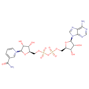
| HET Code: | NAD |
|---|---|
| Formula: | C21H26N7O14P2 |
| Molecular weight: | 662.417 g/mol |
| DrugBank ID: | - |
| Buried Surface Area: | 67.04 % |
| Polar Surface area: | 343.54 Å2 |
| Number of | |
|---|---|
| H-Bond Acceptors: | 18 |
| H-Bond Donors: | 6 |
| Rings: | 5 |
| Aromatic rings: | 3 |
| Anionic atoms: | 2 |
| Cationic atoms: | 1 |
| Rule of Five Violation: | 3 |
| Rotatable Bonds: | 11 |
| X | Y | Z |
|---|---|---|
| -20.0874 | -48.6924 | -25.0884 |
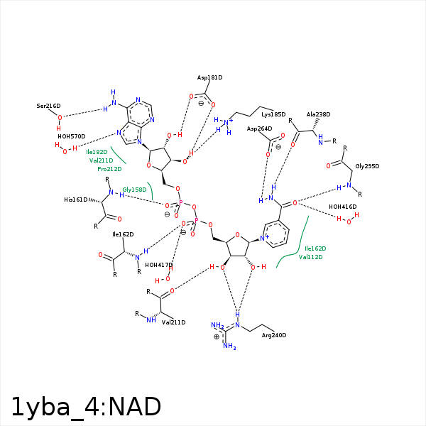


Represent the protein/ligand binding mode, centered on the ligand
Dashed lines represents hydrogen bonds and metal interactions
Green residue labels for amino acids with hydrophobic contacts (green lines) to the ligand
| Ligand | Protein | Interaction | |||
|---|---|---|---|---|---|
| Atom | Atom | Residue | Distance (Å) | Angle (°) | Type |
| C4N | CG2 | VAL- 112 | 3.72 | 0 | Hydrophobic |
| O2A | N | HIS- 161 | 2.89 | 177 | H-Bond (Protein Donor) |
| O2N | N | ILE- 162 | 2.91 | 176.48 | H-Bond (Protein Donor) |
| C5D | CD1 | ILE- 162 | 4.31 | 0 | Hydrophobic |
| C5N | CD1 | ILE- 162 | 3.98 | 0 | Hydrophobic |
| O3B | OD2 | ASP- 181 | 2.65 | 144.75 | H-Bond (Ligand Donor) |
| O2B | OD2 | ASP- 181 | 3.33 | 128.44 | H-Bond (Ligand Donor) |
| O3B | NZ | LYS- 185 | 2.79 | 172.33 | H-Bond (Protein Donor) |
| C5D | CB | HIS- 210 | 4.45 | 0 | Hydrophobic |
| O3D | O | VAL- 211 | 2.61 | 156.85 | H-Bond (Ligand Donor) |
| N6A | OG | SER- 216 | 3.06 | 149.78 | H-Bond (Ligand Donor) |
| N7N | O | ALA- 238 | 2.96 | 167.83 | H-Bond (Ligand Donor) |
| C4D | CB | SER- 239 | 4.06 | 0 | Hydrophobic |
| C3D | CG | ARG- 240 | 4.41 | 0 | Hydrophobic |
| O3D | NE | ARG- 240 | 2.92 | 127.58 | H-Bond (Protein Donor) |
| O2D | NH2 | ARG- 240 | 3.32 | 135.55 | H-Bond (Protein Donor) |
| O2D | NE | ARG- 240 | 3.07 | 149.42 | H-Bond (Protein Donor) |
| N7N | OD2 | ASP- 264 | 3 | 175.49 | H-Bond (Ligand Donor) |
| O7N | N | GLY- 295 | 3.16 | 142.57 | H-Bond (Protein Donor) |
| O7N | O | HOH- 416 | 2.85 | 179.95 | H-Bond (Protein Donor) |
| O2N | O | HOH- 417 | 2.81 | 165.12 | H-Bond (Protein Donor) |
| N7A | O | HOH- 570 | 2.85 | 143.43 | H-Bond (Protein Donor) |
