sc-PDB
An Annotated Database of Druggable Binding Sites from the Protein DataBank
An Annotated Database of Druggable Binding Sites from the Protein DataBank
1.900 Å
X-ray
2004-09-23
| Name: | Vitamin B12-dependent ribonucleotide reductase |
|---|---|
| ID: | O33839_THEMT |
| AC: | O33839 |
| Organism: | Thermotoga maritima |
| Reign: | Bacteria |
| TaxID: | 2336 |
| EC Number: | / |
| Chain Name: | Percentage of Residues within binding site |
|---|---|
| A | 74 % |
| B | 26 % |
| B-Factor: | 31.557 |
|---|---|
| Number of residues: | 33 |
| Including | |
| Standard Amino Acids: | 30 |
| Non Standard Amino Acids: | 1 |
| Water Molecules: | 2 |
| Cofactors: | |
| Metals: | MG |
| Ligandability | Volume (Å3) |
|---|---|
| 0.024 | 313.875 |
| % Hydrophobic | % Polar |
|---|---|
| 50.54 | 49.46 |
| According to VolSite | |
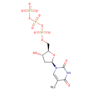
| HET Code: | TTP |
|---|---|
| Formula: | C10H13N2O14P3 |
| Molecular weight: | 478.137 g/mol |
| DrugBank ID: | DB02452 |
| Buried Surface Area: | 68.89 % |
| Polar Surface area: | 279.44 Å2 |
| Number of | |
|---|---|
| H-Bond Acceptors: | 14 |
| H-Bond Donors: | 2 |
| Rings: | 2 |
| Aromatic rings: | 0 |
| Anionic atoms: | 4 |
| Cationic atoms: | 0 |
| Rule of Five Violation: | 1 |
| Rotatable Bonds: | 8 |
| X | Y | Z |
|---|---|---|
| -86.5898 | -37.2588 | -21.147 |
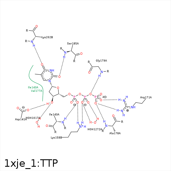


Represent the protein/ligand binding mode, centered on the ligand
Dashed lines represents hydrogen bonds and metal interactions
Green residue labels for amino acids with hydrophobic contacts (green lines) to the ligand
| Ligand | Protein | Interaction | |||
|---|---|---|---|---|---|
| Atom | Atom | Residue | Distance (Å) | Angle (°) | Type |
| O3' | OD1 | ASP- 141 | 2.69 | 173.63 | H-Bond (Ligand Donor) |
| O2A | N | ILE- 143 | 2.94 | 174.36 | H-Bond (Protein Donor) |
| C2' | CD1 | ILE- 143 | 3.82 | 0 | Hydrophobic |
| C5M | CG1 | ILE- 143 | 3.74 | 0 | Hydrophobic |
| C2' | CD1 | ILE- 146 | 3.78 | 0 | Hydrophobic |
| O2A | NZ | LYS- 158 | 2.87 | 169.39 | H-Bond (Protein Donor) |
| O1B | NZ | LYS- 158 | 3.09 | 128 | H-Bond (Protein Donor) |
| O2A | NZ | LYS- 158 | 2.87 | 0 | Ionic (Protein Cationic) |
| O1B | NZ | LYS- 158 | 3.09 | 0 | Ionic (Protein Cationic) |
| O2B | NZ | LYS- 158 | 3.82 | 0 | Ionic (Protein Cationic) |
| C5M | CD | LYS- 158 | 3.84 | 0 | Hydrophobic |
| O1G | NH1 | ARG- 171 | 2.87 | 169.86 | H-Bond (Protein Donor) |
| O2G | NH2 | ARG- 171 | 2.97 | 147.75 | H-Bond (Protein Donor) |
| O1G | CZ | ARG- 171 | 3.78 | 0 | Ionic (Protein Cationic) |
| O2G | CZ | ARG- 171 | 3.69 | 0 | Ionic (Protein Cationic) |
| C4' | CD | ARG- 171 | 3.99 | 0 | Hydrophobic |
| C1' | CG2 | VAL- 177 | 4.34 | 0 | Hydrophobic |
| C5M | CG1 | VAL- 177 | 4.02 | 0 | Hydrophobic |
| O1G | N | ALA- 178 | 2.9 | 160.92 | H-Bond (Protein Donor) |
| O3B | N | GLY- 179 | 3.13 | 157.93 | H-Bond (Protein Donor) |
| C5M | CG2 | THR- 180 | 4.06 | 0 | Hydrophobic |
| C1' | CB | ALA- 184 | 4.13 | 0 | Hydrophobic |
| O2 | N | SER- 185 | 2.69 | 166.01 | H-Bond (Protein Donor) |
| C2' | CZ | PHE- 190 | 3.96 | 0 | Hydrophobic |
| C1' | CE2 | PHE- 190 | 4.12 | 0 | Hydrophobic |
| O4 | N | LYS- 202 | 2.96 | 170.66 | H-Bond (Protein Donor) |
| O1A | MG | MG- 1006 | 2 | 0 | Metal Acceptor |
| O2B | MG | MG- 1006 | 1.94 | 0 | Metal Acceptor |
| O2G | MG | MG- 1006 | 1.91 | 0 | Metal Acceptor |
| O3' | O | HOH- 1017 | 3.16 | 179.97 | H-Bond (Protein Donor) |
| O2B | O | HOH- 1272 | 2.51 | 138.56 | H-Bond (Protein Donor) |
