sc-PDB
An Annotated Database of Druggable Binding Sites from the Protein DataBank
An Annotated Database of Druggable Binding Sites from the Protein DataBank
1.700 Å
X-ray
1994-03-07
| Name: | Xylose isomerase |
|---|---|
| ID: | XYLA_STRRU |
| AC: | P24300 |
| Organism: | Streptomyces rubiginosus |
| Reign: | Bacteria |
| TaxID: | 1929 |
| EC Number: | 5.3.1.5 |
| Chain Name: | Percentage of Residues within binding site |
|---|---|
| A | 100 % |
| B-Factor: | 5.025 |
|---|---|
| Number of residues: | 25 |
| Including | |
| Standard Amino Acids: | 23 |
| Non Standard Amino Acids: | 2 |
| Water Molecules: | 0 |
| Cofactors: | |
| Metals: | MN MN |
| Ligandability | Volume (Å3) |
|---|---|
| 0.373 | 766.125 |
| % Hydrophobic | % Polar |
|---|---|
| 33.48 | 66.52 |
| According to VolSite | |
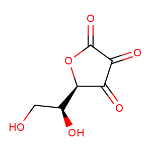
| HET Code: | ASC |
|---|---|
| Formula: | C6H7O6 |
| Molecular weight: | 175.116 g/mol |
| DrugBank ID: | DB00126 |
| Buried Surface Area: | 65.17 % |
| Polar Surface area: | 110.05 Å2 |
| Number of | |
|---|---|
| H-Bond Acceptors: | 6 |
| H-Bond Donors: | 3 |
| Rings: | 1 |
| Aromatic rings: | 0 |
| Anionic atoms: | 1 |
| Cationic atoms: | 0 |
| Rule of Five Violation: | 0 |
| Rotatable Bonds: | 2 |
| X | Y | Z |
|---|---|---|
| 35.4914 | 32.1093 | 57.5571 |
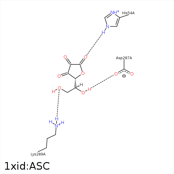


Represent the protein/ligand binding mode, centered on the ligand
Dashed lines represents hydrogen bonds and metal interactions
Green residue labels for amino acids with hydrophobic contacts (green lines) to the ligand
| Ligand | Protein | Interaction | |||
|---|---|---|---|---|---|
| Atom | Atom | Residue | Distance (Å) | Angle (°) | Type |
| O1 | NE2 | HIS- 54 | 2.78 | 147.35 | H-Bond (Protein Donor) |
| O4 | NE2 | HIS- 54 | 3.48 | 142.61 | H-Bond (Protein Donor) |
| C6 | CZ | PHE- 94 | 4.05 | 0 | Hydrophobic |
| C4 | CE3 | TRP- 137 | 3.47 | 0 | Hydrophobic |
| C5 | CZ3 | TRP- 137 | 4.49 | 0 | Hydrophobic |
| O5 | OD2 | ASP- 287 | 2.94 | 125.48 | H-Bond (Ligand Donor) |
| O6 | NZ | LYS- 289 | 2.77 | 165.3 | H-Bond (Protein Donor) |
| O2 | MN | MN- 391 | 2.36 | 0 | Metal Acceptor |
| O3 | MN | MN- 391 | 2.49 | 0 | Metal Acceptor |
