sc-PDB
An Annotated Database of Druggable Binding Sites from the Protein DataBank
An Annotated Database of Druggable Binding Sites from the Protein DataBank
1.650 Å
X-ray
1997-01-06
| Name: | UDP-glucose 4-epimerase |
|---|---|
| ID: | GALE_ECOLI |
| AC: | P09147 |
| Organism: | Escherichia coli |
| Reign: | Bacteria |
| TaxID: | 83333 |
| EC Number: | 5.1.3.2 |
| Chain Name: | Percentage of Residues within binding site |
|---|---|
| A | 100 % |
| B-Factor: | 15.003 |
|---|---|
| Number of residues: | 42 |
| Including | |
| Standard Amino Acids: | 38 |
| Non Standard Amino Acids: | 1 |
| Water Molecules: | 3 |
| Cofactors: | NAD |
| Metals: | |
| Ligandability | Volume (Å3) |
|---|---|
| 0.945 | 1599.750 |
| % Hydrophobic | % Polar |
|---|---|
| 40.93 | 59.07 |
| According to VolSite | |
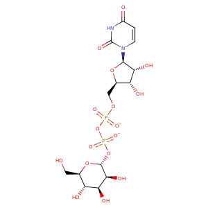
| HET Code: | UFM |
|---|---|
| Formula: | C15H22N2O17P2 |
| Molecular weight: | 564.286 g/mol |
| DrugBank ID: | DB02421 |
| Buried Surface Area: | 67.44 % |
| Polar Surface area: | 316.82 Å2 |
| Number of | |
|---|---|
| H-Bond Acceptors: | 17 |
| H-Bond Donors: | 7 |
| Rings: | 3 |
| Aromatic rings: | 0 |
| Anionic atoms: | 2 |
| Cationic atoms: | 0 |
| Rule of Five Violation: | 3 |
| Rotatable Bonds: | 9 |
| X | Y | Z |
|---|---|---|
| 17.0076 | 11.1852 | 37.3985 |
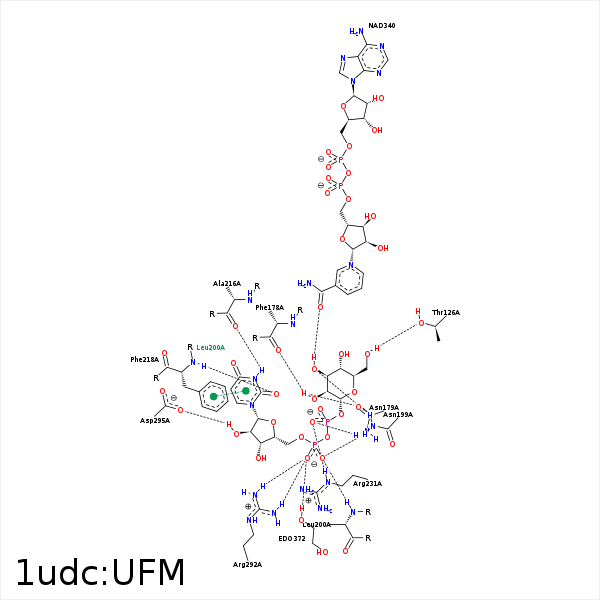


Represent the protein/ligand binding mode, centered on the ligand
Dashed lines represents hydrogen bonds and metal interactions
Green residue labels for amino acids with hydrophobic contacts (green lines) to the ligand
| Ligand | Protein | Interaction | |||
|---|---|---|---|---|---|
| Atom | Atom | Residue | Distance (Å) | Angle (°) | Type |
| C6' | CG2 | VAL- 86 | 4.25 | 0 | Hydrophobic |
| C6' | CB | THR- 126 | 3.66 | 0 | Hydrophobic |
| O6' | OG1 | THR- 126 | 3.12 | 169.15 | H-Bond (Ligand Donor) |
| C4' | CE2 | TYR- 149 | 4.23 | 0 | Hydrophobic |
| C6' | CE2 | TYR- 149 | 3.89 | 0 | Hydrophobic |
| O2' | O | PHE- 178 | 2.94 | 120.88 | H-Bond (Ligand Donor) |
| O1B | ND2 | ASN- 179 | 3 | 158.05 | H-Bond (Protein Donor) |
| O2' | OD1 | ASN- 179 | 3.07 | 136.01 | H-Bond (Ligand Donor) |
| O2A | ND2 | ASN- 199 | 3.01 | 169.93 | H-Bond (Protein Donor) |
| O3' | ND2 | ASN- 199 | 3.02 | 156.17 | H-Bond (Protein Donor) |
| C1D | CD1 | LEU- 200 | 4.36 | 0 | Hydrophobic |
| C4D | CD2 | LEU- 200 | 4.37 | 0 | Hydrophobic |
| C5D | CB | LEU- 200 | 4.15 | 0 | Hydrophobic |
| O2A | N | LEU- 200 | 3.13 | 167.07 | H-Bond (Protein Donor) |
| N3 | O | ALA- 216 | 2.76 | 168.83 | H-Bond (Ligand Donor) |
| O2 | N | PHE- 218 | 2.85 | 161.55 | H-Bond (Protein Donor) |
| O1B | NE | ARG- 231 | 2.89 | 138.28 | H-Bond (Protein Donor) |
| C4D | CG | ARG- 231 | 4.04 | 0 | Hydrophobic |
| C5D | CZ | TYR- 233 | 4.25 | 0 | Hydrophobic |
| C1D | CG2 | VAL- 269 | 3.86 | 0 | Hydrophobic |
| C4D | CG2 | VAL- 269 | 4.35 | 0 | Hydrophobic |
| O5D | NH2 | ARG- 292 | 3.47 | 138.95 | H-Bond (Protein Donor) |
| O1A | NH2 | ARG- 292 | 3.16 | 141.11 | H-Bond (Protein Donor) |
| O1A | NH1 | ARG- 292 | 2.98 | 152.13 | H-Bond (Protein Donor) |
| O1A | CZ | ARG- 292 | 3.49 | 0 | Ionic (Protein Cationic) |
| O2B | CZ | ARG- 292 | 3.73 | 0 | Ionic (Protein Cationic) |
| O2D | OD2 | ASP- 295 | 2.76 | 150.75 | H-Bond (Ligand Donor) |
| O3' | O7N | NAD- 340 | 2.64 | 120.98 | H-Bond (Ligand Donor) |
| C4' | C4N | NAD- 340 | 4.16 | 0 | Hydrophobic |
| O3' | O | HOH- 517 | 3.3 | 143.82 | H-Bond (Protein Donor) |
