sc-PDB
An Annotated Database of Druggable Binding Sites from the Protein DataBank
An Annotated Database of Druggable Binding Sites from the Protein DataBank
1.800 Å
X-ray
1997-01-06
| Name: | UDP-glucose 4-epimerase |
|---|---|
| ID: | GALE_ECOLI |
| AC: | P09147 |
| Organism: | Escherichia coli |
| Reign: | Bacteria |
| TaxID: | 83333 |
| EC Number: | 5.1.3.2 |
| Chain Name: | Percentage of Residues within binding site |
|---|---|
| A | 100 % |
| B-Factor: | 19.336 |
|---|---|
| Number of residues: | 47 |
| Including | |
| Standard Amino Acids: | 41 |
| Non Standard Amino Acids: | 1 |
| Water Molecules: | 5 |
| Cofactors: | NAD |
| Metals: | |
| Ligandability | Volume (Å3) |
|---|---|
| 0.859 | 654.750 |
| % Hydrophobic | % Polar |
|---|---|
| 44.85 | 55.15 |
| According to VolSite | |
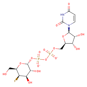
| HET Code: | UFG |
|---|---|
| Formula: | C15H21FN2O16P2 |
| Molecular weight: | 566.277 g/mol |
| DrugBank ID: | DB04097 |
| Buried Surface Area: | 63.57 % |
| Polar Surface area: | 296.59 Å2 |
| Number of | |
|---|---|
| H-Bond Acceptors: | 16 |
| H-Bond Donors: | 6 |
| Rings: | 3 |
| Aromatic rings: | 0 |
| Anionic atoms: | 2 |
| Cationic atoms: | 0 |
| Rule of Five Violation: | 3 |
| Rotatable Bonds: | 9 |
| X | Y | Z |
|---|---|---|
| 16.8458 | 11.3892 | 37.1609 |
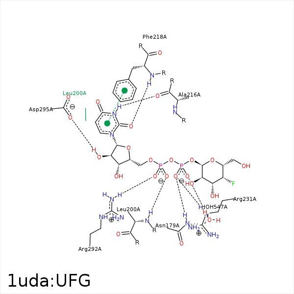


Represent the protein/ligand binding mode, centered on the ligand
Dashed lines represents hydrogen bonds and metal interactions
Green residue labels for amino acids with hydrophobic contacts (green lines) to the ligand
| Ligand | Protein | Interaction | |||
|---|---|---|---|---|---|
| Atom | Atom | Residue | Distance (Å) | Angle (°) | Type |
| C4' | CG2 | VAL- 86 | 4 | 0 | Hydrophobic |
| O3' | OG | SER- 124 | 3.32 | 126.53 | H-Bond (Ligand Donor) |
| C4' | CB | THR- 126 | 4.36 | 0 | Hydrophobic |
| F4' | CG2 | THR- 126 | 4.19 | 0 | Hydrophobic |
| O1B | ND2 | ASN- 179 | 2.87 | 174.11 | H-Bond (Protein Donor) |
| C1D | CD1 | LEU- 200 | 4.42 | 0 | Hydrophobic |
| C4D | CD2 | LEU- 200 | 4.31 | 0 | Hydrophobic |
| C5D | CB | LEU- 200 | 4.1 | 0 | Hydrophobic |
| O2A | N | LEU- 200 | 2.81 | 169.04 | H-Bond (Protein Donor) |
| N3 | O | ALA- 216 | 2.8 | 163.28 | H-Bond (Ligand Donor) |
| O2 | N | PHE- 218 | 2.87 | 165.71 | H-Bond (Protein Donor) |
| C2D | CD2 | PHE- 218 | 4.5 | 0 | Hydrophobic |
| O1B | NE | ARG- 231 | 2.8 | 141.63 | H-Bond (Protein Donor) |
| O1B | CZ | ARG- 231 | 3.83 | 0 | Ionic (Protein Cationic) |
| C4D | CG | ARG- 231 | 4.05 | 0 | Hydrophobic |
| C5D | CZ | TYR- 233 | 4.38 | 0 | Hydrophobic |
| C1D | CG2 | VAL- 269 | 3.83 | 0 | Hydrophobic |
| C4D | CG2 | VAL- 269 | 4.42 | 0 | Hydrophobic |
| O5D | NH2 | ARG- 292 | 3.39 | 137.69 | H-Bond (Protein Donor) |
| O1A | NH2 | ARG- 292 | 2.89 | 156.33 | H-Bond (Protein Donor) |
| O1A | NH1 | ARG- 292 | 3.27 | 136.99 | H-Bond (Protein Donor) |
| O1A | CZ | ARG- 292 | 3.53 | 0 | Ionic (Protein Cationic) |
| O2D | OD2 | ASP- 295 | 2.62 | 157.35 | H-Bond (Ligand Donor) |
| O2B | O | HOH- 547 | 2.59 | 160.44 | H-Bond (Protein Donor) |
