sc-PDB
An Annotated Database of Druggable Binding Sites from the Protein DataBank
An Annotated Database of Druggable Binding Sites from the Protein DataBank
1.650 Å
X-ray
2004-07-23
| Name: | Alcohol dehydrogenase 1B |
|---|---|
| ID: | ADH1B_HUMAN |
| AC: | P00325 |
| Organism: | Homo sapiens |
| Reign: | Eukaryota |
| TaxID: | 9606 |
| EC Number: | 1.1.1.1 |
| Chain Name: | Percentage of Residues within binding site |
|---|---|
| A | 98 % |
| B | 2 % |
| B-Factor: | 10.910 |
|---|---|
| Number of residues: | 56 |
| Including | |
| Standard Amino Acids: | 50 |
| Non Standard Amino Acids: | 1 |
| Water Molecules: | 5 |
| Cofactors: | |
| Metals: | ZN |
| Ligandability | Volume (Å3) |
|---|---|
| 1.393 | 1039.500 |
| % Hydrophobic | % Polar |
|---|---|
| 53.57 | 46.43 |
| According to VolSite | |
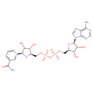
| HET Code: | NAD |
|---|---|
| Formula: | C21H26N7O14P2 |
| Molecular weight: | 662.417 g/mol |
| DrugBank ID: | - |
| Buried Surface Area: | 72.13 % |
| Polar Surface area: | 343.54 Å2 |
| Number of | |
|---|---|
| H-Bond Acceptors: | 18 |
| H-Bond Donors: | 6 |
| Rings: | 5 |
| Aromatic rings: | 3 |
| Anionic atoms: | 2 |
| Cationic atoms: | 1 |
| Rule of Five Violation: | 3 |
| Rotatable Bonds: | 11 |
| X | Y | Z |
|---|---|---|
| 2.76405 | -11.8735 | -16.221 |


Represent the protein/ligand binding mode, centered on the ligand
Dashed lines represents hydrogen bonds and metal interactions
Green residue labels for amino acids with hydrophobic contacts (green lines) to the ligand
| Ligand | Protein | Interaction | |||
|---|---|---|---|---|---|
| Atom | Atom | Residue | Distance (Å) | Angle (°) | Type |
| C5N | SG | CYS- 46 | 4.01 | 0 | Hydrophobic |
| O1A | NE | ARG- 47 | 2.73 | 152.84 | H-Bond (Protein Donor) |
| O1A | NH2 | ARG- 47 | 3 | 136.92 | H-Bond (Protein Donor) |
| O1N | N | ARG- 47 | 3.11 | 159.52 | H-Bond (Protein Donor) |
| O1A | CZ | ARG- 47 | 3.29 | 0 | Ionic (Protein Cationic) |
| C3D | CG | ARG- 47 | 3.89 | 0 | Hydrophobic |
| C2D | CB | ARG- 47 | 4.11 | 0 | Hydrophobic |
| O2D | OG1 | THR- 48 | 2.78 | 158.87 | H-Bond (Ligand Donor) |
| O3D | NE2 | HIS- 51 | 3.02 | 169.63 | H-Bond (Ligand Donor) |
| C5N | SG | CYS- 174 | 3.52 | 0 | Hydrophobic |
| C4N | CG2 | THR- 178 | 3.4 | 0 | Hydrophobic |
| O2N | N | VAL- 203 | 3.12 | 160.36 | H-Bond (Protein Donor) |
| C5D | CB | VAL- 203 | 4.27 | 0 | Hydrophobic |
| C5N | CG2 | VAL- 203 | 4 | 0 | Hydrophobic |
| O3B | OD1 | ASP- 223 | 2.71 | 168.95 | H-Bond (Ligand Donor) |
| O2B | OD2 | ASP- 223 | 2.68 | 174.23 | H-Bond (Ligand Donor) |
| O3B | NZ | LYS- 228 | 2.85 | 136.12 | H-Bond (Protein Donor) |
| C5D | CG1 | VAL- 268 | 4.2 | 0 | Hydrophobic |
| C1B | CG1 | ILE- 269 | 4.28 | 0 | Hydrophobic |
| C3N | CG1 | VAL- 292 | 4.44 | 0 | Hydrophobic |
| N7N | O | VAL- 292 | 3.01 | 176.83 | H-Bond (Ligand Donor) |
| O3D | N | VAL- 294 | 3.04 | 160.73 | H-Bond (Protein Donor) |
| C2D | CG2 | VAL- 294 | 4.35 | 0 | Hydrophobic |
| N7N | O | ALA- 317 | 3.11 | 153.66 | H-Bond (Ligand Donor) |
| O7N | N | TYR- 319 | 2.9 | 161.27 | H-Bond (Protein Donor) |
| O1N | CZ | ARG- 369 | 3.78 | 0 | Ionic (Protein Cationic) |
| O1N | NH1 | ARG- 369 | 2.74 | 152.26 | H-Bond (Protein Donor) |
| O2N | O | HOH- 1387 | 2.74 | 166.12 | H-Bond (Protein Donor) |
| O2A | O | HOH- 1416 | 2.68 | 179.98 | H-Bond (Protein Donor) |
| O2B | O | HOH- 1466 | 2.83 | 179.99 | H-Bond (Protein Donor) |
