sc-PDB
An Annotated Database of Druggable Binding Sites from the Protein DataBank
An Annotated Database of Druggable Binding Sites from the Protein DataBank
1.950 Å
X-ray
2004-07-20
| Name: | Pancreatic alpha-amylase |
|---|---|
| ID: | AMYP_HUMAN |
| AC: | P04746 |
| Organism: | Homo sapiens |
| Reign: | Eukaryota |
| TaxID: | 9606 |
| EC Number: | / |
| Chain Name: | Percentage of Residues within binding site |
|---|---|
| A | 100 % |
| B-Factor: | 21.427 |
|---|---|
| Number of residues: | 25 |
| Including | |
| Standard Amino Acids: | 20 |
| Non Standard Amino Acids: | 0 |
| Water Molecules: | 5 |
| Cofactors: | |
| Metals: | |
| Ligandability | Volume (Å3) |
|---|---|
| 0.771 | 459.000 |
| % Hydrophobic | % Polar |
|---|---|
| 35.29 | 64.71 |
| According to VolSite | |
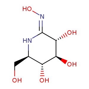
| HET Code: | GOX |
|---|---|
| Formula: | C6H13N2O5 |
| Molecular weight: | 193.178 g/mol |
| DrugBank ID: | DB02376 |
| Buried Surface Area: | 45.28 % |
| Polar Surface area: | 127.15 Å2 |
| Number of | |
|---|---|
| H-Bond Acceptors: | 5 |
| H-Bond Donors: | 7 |
| Rings: | 1 |
| Aromatic rings: | 0 |
| Anionic atoms: | 0 |
| Cationic atoms: | 1 |
| Rule of Five Violation: | 1 |
| Rotatable Bonds: | 1 |
| X | Y | Z |
|---|---|---|
| 7.88069 | 15.8831 | 43.0157 |
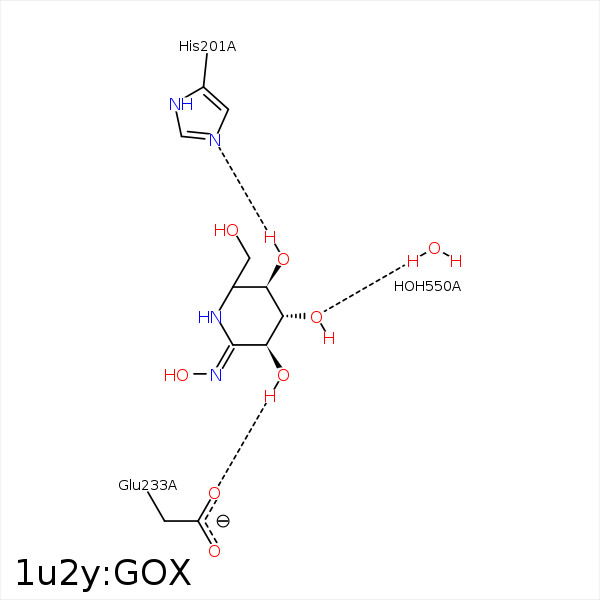


Represent the protein/ligand binding mode, centered on the ligand
Dashed lines represents hydrogen bonds and metal interactions
Green residue labels for amino acids with hydrophobic contacts (green lines) to the ligand
| Ligand | Protein | Interaction | |||
|---|---|---|---|---|---|
| Atom | Atom | Residue | Distance (Å) | Angle (°) | Type |
| C3 | CD2 | LEU- 162 | 4.33 | 0 | Hydrophobic |
| C5 | CD1 | LEU- 162 | 3.84 | 0 | Hydrophobic |
| C3 | CB | ALA- 198 | 4.49 | 0 | Hydrophobic |
| O4 | NE2 | HIS- 201 | 3.15 | 155.22 | H-Bond (Ligand Donor) |
| O2 | OE1 | GLU- 233 | 2.77 | 174.66 | H-Bond (Ligand Donor) |
| C4 | CD1 | ILE- 235 | 3.87 | 0 | Hydrophobic |
| O3 | O | HOH- 550 | 2.56 | 161.58 | H-Bond (Protein Donor) |
