sc-PDB
An Annotated Database of Druggable Binding Sites from the Protein DataBank
An Annotated Database of Druggable Binding Sites from the Protein DataBank
2.700 Å
X-ray
2004-05-22
| Name: | L-amino-acid oxidase |
|---|---|
| ID: | OXLA_GLOHA |
| AC: | Q6STF1 |
| Organism: | Gloydius halys |
| Reign: | Eukaryota |
| TaxID: | 8714 |
| EC Number: | 1.4.3.2 |
| Chain Name: | Percentage of Residues within binding site |
|---|---|
| A | 100 % |
| B-Factor: | 43.828 |
|---|---|
| Number of residues: | 72 |
| Including | |
| Standard Amino Acids: | 69 |
| Non Standard Amino Acids: | 1 |
| Water Molecules: | 2 |
| Cofactors: | |
| Metals: | |
| Ligandability | Volume (Å3) |
|---|---|
| 1.069 | 988.875 |
| % Hydrophobic | % Polar |
|---|---|
| 50.85 | 49.15 |
| According to VolSite | |
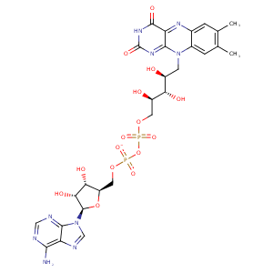
| HET Code: | FAD |
|---|---|
| Formula: | C27H31N9O15P2 |
| Molecular weight: | 783.534 g/mol |
| DrugBank ID: | DB03147 |
| Buried Surface Area: | 77.09 % |
| Polar Surface area: | 381.7 Å2 |
| Number of | |
|---|---|
| H-Bond Acceptors: | 22 |
| H-Bond Donors: | 7 |
| Rings: | 6 |
| Aromatic rings: | 3 |
| Anionic atoms: | 2 |
| Cationic atoms: | 0 |
| Rule of Five Violation: | 3 |
| Rotatable Bonds: | 13 |
| X | Y | Z |
|---|---|---|
| 85.528 | 279.041 | 214.452 |
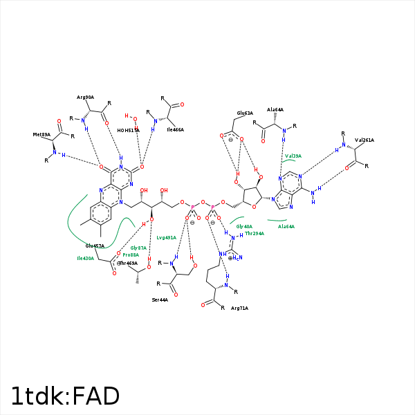


Represent the protein/ligand binding mode, centered on the ligand
Dashed lines represents hydrogen bonds and metal interactions
Green residue labels for amino acids with hydrophobic contacts (green lines) to the ligand
| Ligand | Protein | Interaction | |||
|---|---|---|---|---|---|
| Atom | Atom | Residue | Distance (Å) | Angle (°) | Type |
| O1P | OG | SER- 44 | 2.66 | 152.45 | H-Bond (Protein Donor) |
| O1P | N | SER- 44 | 2.99 | 154.14 | H-Bond (Protein Donor) |
| O3B | OE2 | GLU- 63 | 3.09 | 138.27 | H-Bond (Ligand Donor) |
| O3B | OE1 | GLU- 63 | 3.05 | 161.56 | H-Bond (Ligand Donor) |
| O2B | OE1 | GLU- 63 | 2.56 | 151.13 | H-Bond (Ligand Donor) |
| N3A | N | ALA- 64 | 2.99 | 141.46 | H-Bond (Protein Donor) |
| O1A | N | ARG- 71 | 2.83 | 147.91 | H-Bond (Protein Donor) |
| O2A | NH1 | ARG- 71 | 2.57 | 173.37 | H-Bond (Protein Donor) |
| O2A | CZ | ARG- 71 | 3.44 | 0 | Ionic (Protein Cationic) |
| C9A | CG | PRO- 88 | 4.03 | 0 | Hydrophobic |
| C2' | CG | PRO- 88 | 3.88 | 0 | Hydrophobic |
| O4 | N | MET- 89 | 3.13 | 168.23 | H-Bond (Protein Donor) |
| N3 | O | ARG- 90 | 2.76 | 153.28 | H-Bond (Ligand Donor) |
| O4 | N | ARG- 90 | 3.09 | 163.17 | H-Bond (Protein Donor) |
| N6A | O | VAL- 261 | 3.03 | 157.39 | H-Bond (Ligand Donor) |
| N1A | N | VAL- 261 | 3.01 | 153.75 | H-Bond (Protein Donor) |
| C1B | CG2 | THR- 294 | 4.38 | 0 | Hydrophobic |
| C7M | CE1 | TYR- 372 | 3.95 | 0 | Hydrophobic |
| C8M | CD2 | TRP- 420 | 4.38 | 0 | Hydrophobic |
| C2B | CB | TYR- 425 | 4.04 | 0 | Hydrophobic |
| C7 | CD1 | ILE- 430 | 4.19 | 0 | Hydrophobic |
| C8 | CG1 | ILE- 430 | 3.57 | 0 | Hydrophobic |
| O3' | OE2 | GLU- 457 | 2.97 | 156.23 | H-Bond (Ligand Donor) |
| C5' | CB | GLU- 457 | 3.89 | 0 | Hydrophobic |
| O2P | N | GLU- 457 | 3.41 | 178.31 | H-Bond (Protein Donor) |
| N1 | N | ILE- 466 | 3.44 | 136.08 | H-Bond (Protein Donor) |
| O2 | N | ILE- 466 | 2.81 | 163.96 | H-Bond (Protein Donor) |
| C2' | CG1 | ILE- 466 | 4.21 | 0 | Hydrophobic |
| O3' | OG1 | THR- 469 | 3.06 | 141.98 | H-Bond (Protein Donor) |
| C5' | CG2 | THR- 469 | 3.55 | 0 | Hydrophobic |
| O2 | O | HOH- 517 | 3.14 | 179.96 | H-Bond (Protein Donor) |
