sc-PDB
An Annotated Database of Druggable Binding Sites from the Protein DataBank
An Annotated Database of Druggable Binding Sites from the Protein DataBank
2.700 Å
X-ray
2004-05-19
| Name: | DNA ligase |
|---|---|
| ID: | DNLJ_ENTFA |
| AC: | Q837V6 |
| Organism: | Enterococcus faecalis |
| Reign: | Bacteria |
| TaxID: | 226185 |
| EC Number: | / |
| Chain Name: | Percentage of Residues within binding site |
|---|---|
| A | 100 % |
| B-Factor: | 29.077 |
|---|---|
| Number of residues: | 46 |
| Including | |
| Standard Amino Acids: | 45 |
| Non Standard Amino Acids: | 0 |
| Water Molecules: | 1 |
| Cofactors: | |
| Metals: | |
| Ligandability | Volume (Å3) |
|---|---|
| 0.664 | 1721.250 |
| % Hydrophobic | % Polar |
|---|---|
| 41.76 | 58.24 |
| According to VolSite | |
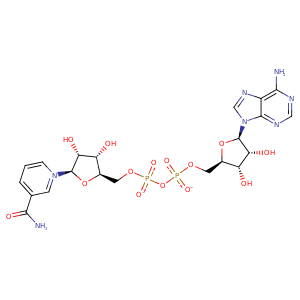
| HET Code: | NAD |
|---|---|
| Formula: | C21H26N7O14P2 |
| Molecular weight: | 662.417 g/mol |
| DrugBank ID: | - |
| Buried Surface Area: | 71.71 % |
| Polar Surface area: | 343.54 Å2 |
| Number of | |
|---|---|
| H-Bond Acceptors: | 18 |
| H-Bond Donors: | 6 |
| Rings: | 5 |
| Aromatic rings: | 3 |
| Anionic atoms: | 2 |
| Cationic atoms: | 1 |
| Rule of Five Violation: | 3 |
| Rotatable Bonds: | 11 |
| X | Y | Z |
|---|---|---|
| -7.91377 | 60.264 | 31.8909 |


Represent the protein/ligand binding mode, centered on the ligand
Dashed lines represents hydrogen bonds and metal interactions
Green residue labels for amino acids with hydrophobic contacts (green lines) to the ligand
| Ligand | Protein | Interaction | |||
|---|---|---|---|---|---|
| Atom | Atom | Residue | Distance (Å) | Angle (°) | Type |
| C4N | CB | TYR- 25 | 4.33 | 0 | Hydrophobic |
| DuAr | DuAr | TYR- 29 | 3.7 | 0 | Aromatic Face/Face |
| C4N | CB | TYR- 29 | 3.4 | 0 | Hydrophobic |
| N7N | O | VAL- 37 | 2.76 | 167.07 | H-Bond (Ligand Donor) |
| N7N | OD2 | ASP- 39 | 2.91 | 151.78 | H-Bond (Ligand Donor) |
| C2D | CD1 | TYR- 42 | 4.22 | 0 | Hydrophobic |
| C3N | CB | TYR- 42 | 3.79 | 0 | Hydrophobic |
| DuAr | DuAr | TYR- 42 | 3.79 | 0 | Aromatic Face/Face |
| O2D | OD2 | ASP- 43 | 3.15 | 127.62 | H-Bond (Ligand Donor) |
| O2D | OD1 | ASP- 43 | 2.72 | 167.34 | H-Bond (Ligand Donor) |
| O3 | OG | SER- 88 | 3.16 | 121.39 | H-Bond (Protein Donor) |
| O1N | OG | SER- 88 | 2.73 | 152.96 | H-Bond (Protein Donor) |
| C5B | CD1 | LEU- 89 | 3.48 | 0 | Hydrophobic |
| O2A | OD2 | ASP- 91 | 2.54 | 169.62 | H-Bond (Protein Donor) |
| N6A | OE2 | GLU- 118 | 2.94 | 151.79 | H-Bond (Ligand Donor) |
| O1A | NZ | LYS- 120 | 3.17 | 146.97 | H-Bond (Protein Donor) |
| O4B | NZ | LYS- 120 | 3.02 | 138.33 | H-Bond (Protein Donor) |
| O1A | NZ | LYS- 120 | 3.17 | 0 | Ionic (Protein Cationic) |
| C1B | CG | LYS- 120 | 4.46 | 0 | Hydrophobic |
| N7A | N | ILE- 121 | 3.02 | 131.09 | H-Bond (Protein Donor) |
| O1N | NH1 | ARG- 141 | 3.09 | 134.95 | H-Bond (Protein Donor) |
| O1N | NH2 | ARG- 141 | 2.71 | 157.29 | H-Bond (Protein Donor) |
| O1N | CZ | ARG- 141 | 3.32 | 0 | Ionic (Protein Cationic) |
| O2B | OE2 | GLU- 175 | 2.71 | 169.89 | H-Bond (Ligand Donor) |
| C2B | CZ | TYR- 227 | 4.42 | 0 | Hydrophobic |
| DuAr | DuAr | TYR- 227 | 3.62 | 0 | Aromatic Face/Face |
| N1A | NZ | LYS- 291 | 2.81 | 148.34 | H-Bond (Protein Donor) |
| O1A | NZ | LYS- 315 | 3.13 | 125.89 | H-Bond (Protein Donor) |
| O3D | NZ | LYS- 315 | 2.86 | 122.37 | H-Bond (Protein Donor) |
| O1A | NZ | LYS- 315 | 3.13 | 0 | Ionic (Protein Cationic) |
