sc-PDB
An Annotated Database of Druggable Binding Sites from the Protein DataBank
An Annotated Database of Druggable Binding Sites from the Protein DataBank
1.900 Å
X-ray
2004-03-24
| Name: | m7GpppX diphosphatase |
|---|---|
| ID: | DCPS_HUMAN |
| AC: | Q96C86 |
| Organism: | Homo sapiens |
| Reign: | Eukaryota |
| TaxID: | 9606 |
| EC Number: | 3.6.1.59 |
| Chain Name: | Percentage of Residues within binding site |
|---|---|
| A | 86 % |
| B | 14 % |
| B-Factor: | 25.692 |
|---|---|
| Number of residues: | 50 |
| Including | |
| Standard Amino Acids: | 50 |
| Non Standard Amino Acids: | 0 |
| Water Molecules: | 0 |
| Cofactors: | |
| Metals: | |
| Ligandability | Volume (Å3) |
|---|---|
| 0.500 | 1957.500 |
| % Hydrophobic | % Polar |
|---|---|
| 41.90 | 58.10 |
| According to VolSite | |
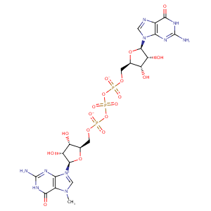
| HET Code: | GTG |
|---|---|
| Formula: | C21H27N10O18P3 |
| Molecular weight: | 800.417 g/mol |
| DrugBank ID: | DB03958 |
| Buried Surface Area: | 72.46 % |
| Polar Surface area: | 447.71 Å2 |
| Number of | |
|---|---|
| H-Bond Acceptors: | 23 |
| H-Bond Donors: | 8 |
| Rings: | 6 |
| Aromatic rings: | 2 |
| Anionic atoms: | 3 |
| Cationic atoms: | 1 |
| Rule of Five Violation: | 3 |
| Rotatable Bonds: | 12 |
| X | Y | Z |
|---|---|---|
| 35.1615 | 57.0563 | 55.8143 |


Represent the protein/ligand binding mode, centered on the ligand
Dashed lines represents hydrogen bonds and metal interactions
Green residue labels for amino acids with hydrophobic contacts (green lines) to the ligand
| Ligand | Protein | Interaction | |||
|---|---|---|---|---|---|
| Atom | Atom | Residue | Distance (Å) | Angle (°) | Type |
| O2E | NH2 | ARG- 54 | 2.93 | 148.66 | H-Bond (Protein Donor) |
| N2B | OD1 | ASP- 59 | 3.04 | 139.95 | H-Bond (Ligand Donor) |
| N1B | OE1 | GLU- 85 | 3.47 | 132.43 | H-Bond (Ligand Donor) |
| N1B | OE2 | GLU- 85 | 2.61 | 156.63 | H-Bond (Ligand Donor) |
| N2B | OE1 | GLU- 85 | 2.83 | 167.46 | H-Bond (Ligand Donor) |
| C7X | CE1 | TYR- 113 | 3.35 | 0 | Hydrophobic |
| O6B | NZ | LYS- 128 | 2.94 | 139.72 | H-Bond (Protein Donor) |
| O1B | NZ | LYS- 142 | 3.65 | 0 | Ionic (Protein Cationic) |
| O2B | NZ | LYS- 142 | 3.39 | 0 | Ionic (Protein Cationic) |
| O1G | NZ | LYS- 142 | 3.88 | 0 | Ionic (Protein Cationic) |
| C7X | CE2 | TRP- 175 | 3.65 | 0 | Hydrophobic |
| C5D | CZ2 | TRP- 175 | 4.44 | 0 | Hydrophobic |
| DuAr | DuAr | TRP- 175 | 3.52 | 0 | Aromatic Face/Face |
| N1A | OE2 | GLU- 185 | 2.7 | 160.95 | H-Bond (Ligand Donor) |
| N2A | OE1 | GLU- 185 | 2.83 | 174.5 | H-Bond (Ligand Donor) |
| N2A | OE2 | GLU- 185 | 3.42 | 129.15 | H-Bond (Ligand Donor) |
| N2A | O | PRO- 204 | 3.04 | 139.46 | H-Bond (Ligand Donor) |
| O3D | OD2 | ASP- 205 | 2.52 | 157.83 | H-Bond (Ligand Donor) |
| O2D | OD1 | ASP- 205 | 2.7 | 149.82 | H-Bond (Ligand Donor) |
| O2D | OD2 | ASP- 205 | 3.32 | 148.23 | H-Bond (Ligand Donor) |
| C7X | CD1 | LEU- 206 | 3.92 | 0 | Hydrophobic |
| C2D | CB | LEU- 206 | 4.38 | 0 | Hydrophobic |
| N3A | N | LEU- 206 | 3.24 | 148.23 | H-Bond (Protein Donor) |
| O2D | NZ | LYS- 207 | 3.31 | 158.46 | H-Bond (Protein Donor) |
| C4D | CD1 | ILE- 219 | 3.79 | 0 | Hydrophobic |
| O2A | N | SER- 272 | 2.87 | 132.51 | H-Bond (Protein Donor) |
| O3A | N | SER- 272 | 3.27 | 160.12 | H-Bond (Protein Donor) |
| O1B | OG | SER- 272 | 2.59 | 148.8 | H-Bond (Protein Donor) |
| O2A | N | TYR- 273 | 2.96 | 162.92 | H-Bond (Protein Donor) |
| C7X | CZ | TYR- 273 | 4.1 | 0 | Hydrophobic |
| C5D | CD1 | TYR- 273 | 3.73 | 0 | Hydrophobic |
| O1A | NE2 | HIS- 279 | 3.2 | 129.18 | H-Bond (Protein Donor) |
| C4E | CB | PRO- 288 | 4.13 | 0 | Hydrophobic |
| C1E | CB | PRO- 288 | 4.26 | 0 | Hydrophobic |
| O3A | NH2 | ARG- 294 | 3.26 | 130.47 | H-Bond (Protein Donor) |
| O1B | NH1 | ARG- 294 | 3.34 | 135.71 | H-Bond (Protein Donor) |
| O1B | NH2 | ARG- 294 | 2.91 | 160 | H-Bond (Protein Donor) |
| O5E | NH1 | ARG- 294 | 3.38 | 142.07 | H-Bond (Protein Donor) |
| O1B | CZ | ARG- 294 | 3.55 | 0 | Ionic (Protein Cationic) |
