sc-PDB
An Annotated Database of Druggable Binding Sites from the Protein DataBank
An Annotated Database of Druggable Binding Sites from the Protein DataBank
2.000 Å
X-ray
1996-09-19
| Name: | Dihydrofolate reductase |
|---|---|
| ID: | DYR_ECOLI |
| AC: | P0ABQ4 |
| Organism: | Escherichia coli |
| Reign: | Bacteria |
| TaxID: | 83333 |
| EC Number: | 1.5.1.3 |
| Chain Name: | Percentage of Residues within binding site |
|---|---|
| A | 100 % |
| B-Factor: | 22.064 |
|---|---|
| Number of residues: | 42 |
| Including | |
| Standard Amino Acids: | 42 |
| Non Standard Amino Acids: | 0 |
| Water Molecules: | 0 |
| Cofactors: | |
| Metals: | |
| Ligandability | Volume (Å3) |
|---|---|
| 1.237 | 590.625 |
| % Hydrophobic | % Polar |
|---|---|
| 53.71 | 46.29 |
| According to VolSite | |
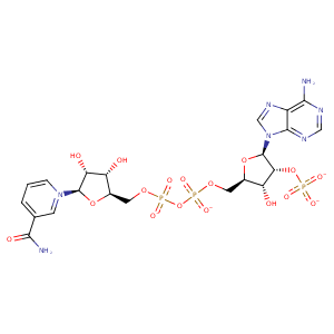
| HET Code: | NAP |
|---|---|
| Formula: | C21H25N7O17P3 |
| Molecular weight: | 740.381 g/mol |
| DrugBank ID: | DB03461 |
| Buried Surface Area: | 68.73 % |
| Polar Surface area: | 405.54 Å2 |
| Number of | |
|---|---|
| H-Bond Acceptors: | 21 |
| H-Bond Donors: | 5 |
| Rings: | 5 |
| Aromatic rings: | 3 |
| Anionic atoms: | 4 |
| Cationic atoms: | 1 |
| Rule of Five Violation: | 2 |
| Rotatable Bonds: | 13 |
| X | Y | Z |
|---|---|---|
| 29.4088 | 54.0245 | 15.0613 |


Represent the protein/ligand binding mode, centered on the ligand
Dashed lines represents hydrogen bonds and metal interactions
Green residue labels for amino acids with hydrophobic contacts (green lines) to the ligand
| Ligand | Protein | Interaction | |||
|---|---|---|---|---|---|
| Atom | Atom | Residue | Distance (Å) | Angle (°) | Type |
| O7N | N | ALA- 7 | 2.68 | 154.83 | H-Bond (Protein Donor) |
| N7N | O | ALA- 7 | 2.87 | 137.38 | H-Bond (Ligand Donor) |
| N7N | O | ILE- 14 | 3.05 | 164.99 | H-Bond (Ligand Donor) |
| C3D | CB | ASN- 18 | 4.26 | 0 | Hydrophobic |
| C3N | SD | MET- 20 | 4.07 | 0 | Hydrophobic |
| C4B | CB | ARG- 44 | 4.32 | 0 | Hydrophobic |
| C1B | CB | ARG- 44 | 4.42 | 0 | Hydrophobic |
| O4B | N | ARG- 44 | 3.13 | 149.3 | H-Bond (Protein Donor) |
| O1X | NE | ARG- 44 | 2.67 | 162.43 | H-Bond (Protein Donor) |
| O3X | NH2 | ARG- 44 | 3.05 | 170.69 | H-Bond (Protein Donor) |
| O1X | CZ | ARG- 44 | 3.64 | 0 | Ionic (Protein Cationic) |
| O3X | CZ | ARG- 44 | 3.85 | 0 | Ionic (Protein Cationic) |
| O5B | N | HIS- 45 | 3.33 | 156.95 | H-Bond (Protein Donor) |
| C5D | CB | HIS- 45 | 4.1 | 0 | Hydrophobic |
| O2A | OG1 | THR- 46 | 2.59 | 154.17 | H-Bond (Protein Donor) |
| O2A | N | THR- 46 | 2.88 | 134.73 | H-Bond (Protein Donor) |
| C5N | CG2 | THR- 46 | 4.15 | 0 | Hydrophobic |
| C1B | CB | LEU- 62 | 4.47 | 0 | Hydrophobic |
| C1B | CB | SER- 63 | 4.49 | 0 | Hydrophobic |
| O1X | OG | SER- 63 | 2.8 | 158.82 | H-Bond (Protein Donor) |
| O2X | OG | SER- 64 | 2.51 | 160.47 | H-Bond (Protein Donor) |
| O2X | N | SER- 64 | 2.93 | 154.18 | H-Bond (Protein Donor) |
| O1A | N | GLY- 96 | 2.98 | 130.22 | H-Bond (Protein Donor) |
| O2A | N | GLY- 96 | 3.03 | 139.72 | H-Bond (Protein Donor) |
| O5D | N | GLY- 97 | 3.35 | 139.03 | H-Bond (Protein Donor) |
| C5B | CB | ARG- 98 | 4.34 | 0 | Hydrophobic |
| O1N | N | ARG- 98 | 2.92 | 176.03 | H-Bond (Protein Donor) |
| O1A | N | VAL- 99 | 3.33 | 151.38 | H-Bond (Protein Donor) |
| C5B | CG2 | VAL- 99 | 4.46 | 0 | Hydrophobic |
| N7A | NE2 | GLN- 102 | 3.13 | 174.66 | H-Bond (Protein Donor) |
| N6A | OE1 | GLN- 102 | 3.1 | 162.96 | H-Bond (Ligand Donor) |
