sc-PDB
An Annotated Database of Druggable Binding Sites from the Protein DataBank
An Annotated Database of Druggable Binding Sites from the Protein DataBank
2.000 Å
X-ray
2003-02-17
| Name: | DNA beta-glucosyltransferase |
|---|---|
| ID: | GSTB_BPT4 |
| AC: | P04547 |
| Organism: | Enterobacteria phage T4 |
| Reign: | Viruses |
| TaxID: | 10665 |
| EC Number: | / |
| Chain Name: | Percentage of Residues within binding site |
|---|---|
| A | 100 % |
| B-Factor: | 23.058 |
|---|---|
| Number of residues: | 51 |
| Including | |
| Standard Amino Acids: | 44 |
| Non Standard Amino Acids: | 1 |
| Water Molecules: | 6 |
| Cofactors: | |
| Metals: | CL |
| Ligandability | Volume (Å3) |
|---|---|
| 0.896 | 661.500 |
| % Hydrophobic | % Polar |
|---|---|
| 44.39 | 55.61 |
| According to VolSite | |
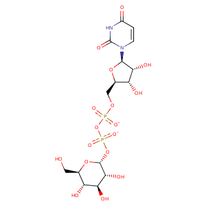
| HET Code: | UPG |
|---|---|
| Formula: | C15H22N2O17P2 |
| Molecular weight: | 564.286 g/mol |
| DrugBank ID: | DB01861 |
| Buried Surface Area: | 74.03 % |
| Polar Surface area: | 316.82 Å2 |
| Number of | |
|---|---|
| H-Bond Acceptors: | 17 |
| H-Bond Donors: | 7 |
| Rings: | 3 |
| Aromatic rings: | 0 |
| Anionic atoms: | 2 |
| Cationic atoms: | 0 |
| Rule of Five Violation: | 3 |
| Rotatable Bonds: | 9 |
| X | Y | Z |
|---|---|---|
| 4.79736 | 1.28928 | 26.0979 |


Represent the protein/ligand binding mode, centered on the ligand
Dashed lines represents hydrogen bonds and metal interactions
Green residue labels for amino acids with hydrophobic contacts (green lines) to the ligand
| Ligand | Protein | Interaction | |||
|---|---|---|---|---|---|
| Atom | Atom | Residue | Distance (Å) | Angle (°) | Type |
| C1C | CG2 | VAL- 18 | 3.87 | 0 | Hydrophobic |
| C4C | CG2 | VAL- 18 | 4.22 | 0 | Hydrophobic |
| C6' | CG1 | VAL- 18 | 4.28 | 0 | Hydrophobic |
| O6' | OE1 | GLU- 22 | 2.76 | 150.85 | H-Bond (Ligand Donor) |
| O3' | O | THR- 99 | 2.64 | 171.51 | H-Bond (Ligand Donor) |
| O4' | NE2 | GLN- 137 | 3.12 | 176.15 | H-Bond (Protein Donor) |
| O1A | N | SER- 189 | 3.01 | 159.6 | H-Bond (Protein Donor) |
| O2A | OG | SER- 189 | 2.63 | 155.96 | H-Bond (Protein Donor) |
| O1B | NH2 | ARG- 191 | 2.85 | 127.76 | H-Bond (Protein Donor) |
| O1B | CZ | ARG- 191 | 3.56 | 0 | Ionic (Protein Cationic) |
| O1B | CZ | ARG- 195 | 3.78 | 0 | Ionic (Protein Cationic) |
| O1B | NH1 | ARG- 195 | 2.94 | 179.35 | H-Bond (Protein Donor) |
| N3 | O | ILE- 238 | 2.96 | 168.1 | H-Bond (Ligand Donor) |
| O4 | N | ILE- 238 | 3 | 148.79 | H-Bond (Protein Donor) |
| C1C | CE | MET- 240 | 4.4 | 0 | Hydrophobic |
| C2C | CG2 | VAL- 243 | 3.74 | 0 | Hydrophobic |
| O2' | OH | TYR- 261 | 2.87 | 148.71 | H-Bond (Ligand Donor) |
| C2' | CE2 | TYR- 261 | 4.13 | 0 | Hydrophobic |
| C3' | CG2 | THR- 267 | 3.71 | 0 | Hydrophobic |
| O4' | N | LEU- 268 | 3.05 | 161.13 | H-Bond (Protein Donor) |
| C6' | CB | LEU- 268 | 3.87 | 0 | Hydrophobic |
| O2B | NE | ARG- 269 | 3.11 | 133.02 | H-Bond (Protein Donor) |
| O2B | NH2 | ARG- 269 | 2.84 | 142.62 | H-Bond (Protein Donor) |
| O2B | CZ | ARG- 269 | 3.38 | 0 | Ionic (Protein Cationic) |
| O2C | OE1 | GLU- 272 | 3.27 | 123.66 | H-Bond (Ligand Donor) |
| O2C | OE2 | GLU- 272 | 2.52 | 157.61 | H-Bond (Ligand Donor) |
| O3C | OE1 | GLU- 272 | 2.52 | 158.49 | H-Bond (Ligand Donor) |
| O2B | O | HOH- 363 | 2.87 | 179.99 | H-Bond (Protein Donor) |
| O4' | O | HOH- 375 | 2.72 | 163.72 | H-Bond (Ligand Donor) |
| O1A | O | HOH- 401 | 2.62 | 179.96 | H-Bond (Protein Donor) |
| O1B | O | HOH- 545 | 2.7 | 162.24 | H-Bond (Protein Donor) |
