sc-PDB
An Annotated Database of Druggable Binding Sites from the Protein DataBank
An Annotated Database of Druggable Binding Sites from the Protein DataBank
1.800 Å
X-ray
1995-11-22
| Name: | UDP-glucose 4-epimerase |
|---|---|
| ID: | GALE_ECOLI |
| AC: | P09147 |
| Organism: | Escherichia coli |
| Reign: | Bacteria |
| TaxID: | 83333 |
| EC Number: | 5.1.3.2 |
| Chain Name: | Percentage of Residues within binding site |
|---|---|
| A | 100 % |
| B-Factor: | 14.455 |
|---|---|
| Number of residues: | 49 |
| Including | |
| Standard Amino Acids: | 46 |
| Non Standard Amino Acids: | 1 |
| Water Molecules: | 2 |
| Cofactors: | |
| Metals: | |
| Ligandability | Volume (Å3) |
|---|---|
| 0.460 | 2025.000 |
| % Hydrophobic | % Polar |
|---|---|
| 41.00 | 59.00 |
| According to VolSite | |
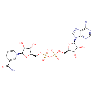
| HET Code: | NAD |
|---|---|
| Formula: | C21H26N7O14P2 |
| Molecular weight: | 662.417 g/mol |
| DrugBank ID: | - |
| Buried Surface Area: | 78.58 % |
| Polar Surface area: | 343.54 Å2 |
| Number of | |
|---|---|
| H-Bond Acceptors: | 18 |
| H-Bond Donors: | 6 |
| Rings: | 5 |
| Aromatic rings: | 3 |
| Anionic atoms: | 2 |
| Cationic atoms: | 1 |
| Rule of Five Violation: | 3 |
| Rotatable Bonds: | 11 |
| X | Y | Z |
|---|---|---|
| 30.122 | 3.04177 | 34.8813 |


Represent the protein/ligand binding mode, centered on the ligand
Dashed lines represents hydrogen bonds and metal interactions
Green residue labels for amino acids with hydrophobic contacts (green lines) to the ligand
| Ligand | Protein | Interaction | |||
|---|---|---|---|---|---|
| Atom | Atom | Residue | Distance (Å) | Angle (°) | Type |
| O2A | N | TYR- 11 | 2.76 | 167.16 | H-Bond (Protein Donor) |
| O2N | N | ILE- 12 | 2.84 | 156.17 | H-Bond (Protein Donor) |
| C5D | CD1 | ILE- 12 | 4.17 | 0 | Hydrophobic |
| O3B | OD2 | ASP- 31 | 2.65 | 164.5 | H-Bond (Ligand Donor) |
| O3B | OD1 | ASP- 31 | 3.18 | 131.14 | H-Bond (Ligand Donor) |
| O2B | OD1 | ASP- 31 | 2.69 | 167.98 | H-Bond (Ligand Donor) |
| N3A | N | ASN- 32 | 3.14 | 150.32 | H-Bond (Protein Donor) |
| C2B | SG | CYS- 34 | 3.82 | 0 | Hydrophobic |
| O1A | ND2 | ASN- 35 | 2.72 | 175.92 | H-Bond (Protein Donor) |
| O2B | N | ASN- 35 | 3.01 | 148.19 | H-Bond (Protein Donor) |
| C2B | CB | ASN- 35 | 3.81 | 0 | Hydrophobic |
| C3B | CB | SER- 36 | 4.37 | 0 | Hydrophobic |
| O3B | OG | SER- 36 | 2.64 | 170.52 | H-Bond (Protein Donor) |
| N6A | OD1 | ASP- 58 | 3.13 | 152.65 | H-Bond (Ligand Donor) |
| N1A | N | ILE- 59 | 3.1 | 172.13 | H-Bond (Protein Donor) |
| C5D | CB | PHE- 80 | 3.83 | 0 | Hydrophobic |
| C1B | CB | ALA- 81 | 4.34 | 0 | Hydrophobic |
| O1N | NZ | LYS- 84 | 2.79 | 171.8 | H-Bond (Protein Donor) |
| O1N | NZ | LYS- 84 | 2.79 | 0 | Ionic (Protein Cationic) |
| C2D | CB | LYS- 84 | 3.94 | 0 | Hydrophobic |
| N6A | OD1 | ASN- 99 | 2.92 | 158.87 | H-Bond (Ligand Donor) |
| C1D | CB | SER- 122 | 3.84 | 0 | Hydrophobic |
| C4D | CB | SER- 122 | 3.68 | 0 | Hydrophobic |
| O2D | OH | TYR- 149 | 2.79 | 159.28 | H-Bond (Ligand Donor) |
| O7N | OH | TYR- 149 | 2.71 | 166.1 | H-Bond (Protein Donor) |
| O3D | NZ | LYS- 153 | 2.79 | 160.39 | H-Bond (Protein Donor) |
| C4D | CE2 | TYR- 177 | 4.36 | 0 | Hydrophobic |
| C1D | CE2 | TYR- 177 | 3.9 | 0 | Hydrophobic |
| C4N | CB | TYR- 177 | 4 | 0 | Hydrophobic |
| N7N | O | PHE- 178 | 3.15 | 153.94 | H-Bond (Ligand Donor) |
| C4N | CG | PRO- 180 | 3.85 | 0 | Hydrophobic |
| O5B | O | HOH- 359 | 3.17 | 162.76 | H-Bond (Protein Donor) |
