sc-PDB
An Annotated Database of Druggable Binding Sites from the Protein DataBank
An Annotated Database of Druggable Binding Sites from the Protein DataBank
2.300 Å
X-ray
2002-10-01
| Name: | Type 2 DNA topoisomerase 6 subunit B |
|---|---|
| ID: | TOP6B_SULSH |
| AC: | O05207 |
| Organism: | Sulfolobus shibatae |
| Reign: | Archaea |
| TaxID: | 2286 |
| EC Number: | / |
| Chain Name: | Percentage of Residues within binding site |
|---|---|
| A | 95 % |
| B | 5 % |
| B-Factor: | 9.327 |
|---|---|
| Number of residues: | 44 |
| Including | |
| Standard Amino Acids: | 42 |
| Non Standard Amino Acids: | 1 |
| Water Molecules: | 1 |
| Cofactors: | |
| Metals: | MG |
| Ligandability | Volume (Å3) |
|---|---|
| 0.339 | 374.625 |
| % Hydrophobic | % Polar |
|---|---|
| 50.45 | 49.55 |
| According to VolSite | |
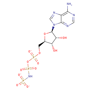
| HET Code: | ANP |
|---|---|
| Formula: | C10H13N6O12P3 |
| Molecular weight: | 502.164 g/mol |
| DrugBank ID: | - |
| Buried Surface Area: | 78.1 % |
| Polar Surface area: | 322.68 Å2 |
| Number of | |
|---|---|
| H-Bond Acceptors: | 16 |
| H-Bond Donors: | 4 |
| Rings: | 3 |
| Aromatic rings: | 2 |
| Anionic atoms: | 4 |
| Cationic atoms: | 0 |
| Rule of Five Violation: | 2 |
| Rotatable Bonds: | 8 |
| X | Y | Z |
|---|---|---|
| 55.5119 | 36.1705 | 25.719 |


Represent the protein/ligand binding mode, centered on the ligand
Dashed lines represents hydrogen bonds and metal interactions
Green residue labels for amino acids with hydrophobic contacts (green lines) to the ligand
| Ligand | Protein | Interaction | |||
|---|---|---|---|---|---|
| Atom | Atom | Residue | Distance (Å) | Angle (°) | Type |
| C1' | CE1 | PHE- 7 | 4.21 | 0 | Hydrophobic |
| O1A | ND2 | ASN- 42 | 2.93 | 148.6 | H-Bond (Protein Donor) |
| N6 | OD1 | ASP- 76 | 3.03 | 152.24 | H-Bond (Ligand Donor) |
| C1' | CD1 | ILE- 81 | 3.76 | 0 | Hydrophobic |
| C4' | CB | ALA- 89 | 4.11 | 0 | Hydrophobic |
| C1' | CB | ALA- 89 | 4.31 | 0 | Hydrophobic |
| O2B | OG | SER- 96 | 2.9 | 172.43 | H-Bond (Protein Donor) |
| O3' | N | SER- 97 | 3.15 | 171.96 | H-Bond (Protein Donor) |
| O2' | OG | SER- 97 | 2.72 | 148.45 | H-Bond (Ligand Donor) |
| O1B | NZ | LYS- 98 | 2.96 | 157.29 | H-Bond (Protein Donor) |
| O1B | NZ | LYS- 98 | 2.96 | 0 | Ionic (Protein Cationic) |
| O2B | NZ | LYS- 98 | 3.87 | 0 | Ionic (Protein Cationic) |
| O3G | N | MET- 107 | 2.94 | 162.81 | H-Bond (Protein Donor) |
| O3G | N | TYR- 108 | 3.16 | 176.2 | H-Bond (Protein Donor) |
| O1G | N | LEU- 110 | 2.65 | 159.03 | H-Bond (Protein Donor) |
| O1G | N | GLY- 111 | 2.58 | 164.14 | H-Bond (Protein Donor) |
| O1A | N | VAL- 112 | 3.26 | 146.57 | H-Bond (Protein Donor) |
| O2A | N | VAL- 112 | 3.27 | 150.23 | H-Bond (Protein Donor) |
| O2A | NZ | LYS- 113 | 2.71 | 150.12 | H-Bond (Protein Donor) |
| O2A | NZ | LYS- 113 | 2.71 | 0 | Ionic (Protein Cationic) |
| O2G | NZ | LYS- 427 | 3.75 | 0 | Ionic (Protein Cationic) |
| O3G | NZ | LYS- 427 | 2.72 | 0 | Ionic (Protein Cationic) |
| O3G | NZ | LYS- 427 | 2.72 | 164.84 | H-Bond (Protein Donor) |
| O2G | MG | MG- 501 | 2.04 | 0 | Metal Acceptor |
| O1B | MG | MG- 501 | 2 | 0 | Metal Acceptor |
| O1A | MG | MG- 501 | 2.1 | 0 | Metal Acceptor |
| N1 | O | HOH- 969 | 2.94 | 179.95 | H-Bond (Protein Donor) |
