sc-PDB
An Annotated Database of Druggable Binding Sites from the Protein DataBank
An Annotated Database of Druggable Binding Sites from the Protein DataBank
2.020 Å
X-ray
2002-09-24
| Name: | GDP-mannose 6-dehydrogenase |
|---|---|
| ID: | ALGD_PSEAE |
| AC: | P11759 |
| Organism: | Pseudomonas aeruginosa |
| Reign: | Bacteria |
| TaxID: | 208964 |
| EC Number: | 1.1.1.132 |
| Chain Name: | Percentage of Residues within binding site |
|---|---|
| A | 51 % |
| B | 49 % |
| B-Factor: | 18.052 |
|---|---|
| Number of residues: | 45 |
| Including | |
| Standard Amino Acids: | 40 |
| Non Standard Amino Acids: | 1 |
| Water Molecules: | 4 |
| Cofactors: | NAD |
| Metals: | |
| Ligandability | Volume (Å3) |
|---|---|
| 0.890 | 624.375 |
| % Hydrophobic | % Polar |
|---|---|
| 47.03 | 52.97 |
| According to VolSite | |
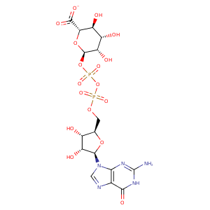
| HET Code: | GDX |
|---|---|
| Formula: | C16H20N5O17P2 |
| Molecular weight: | 616.301 g/mol |
| DrugBank ID: | DB04023 |
| Buried Surface Area: | 73.44 % |
| Polar Surface area: | 372.61 Å2 |
| Number of | |
|---|---|
| H-Bond Acceptors: | 20 |
| H-Bond Donors: | 7 |
| Rings: | 4 |
| Aromatic rings: | 1 |
| Anionic atoms: | 3 |
| Cationic atoms: | 0 |
| Rule of Five Violation: | 3 |
| Rotatable Bonds: | 9 |
| X | Y | Z |
|---|---|---|
| -16.098 | 6.38857 | -11.9987 |
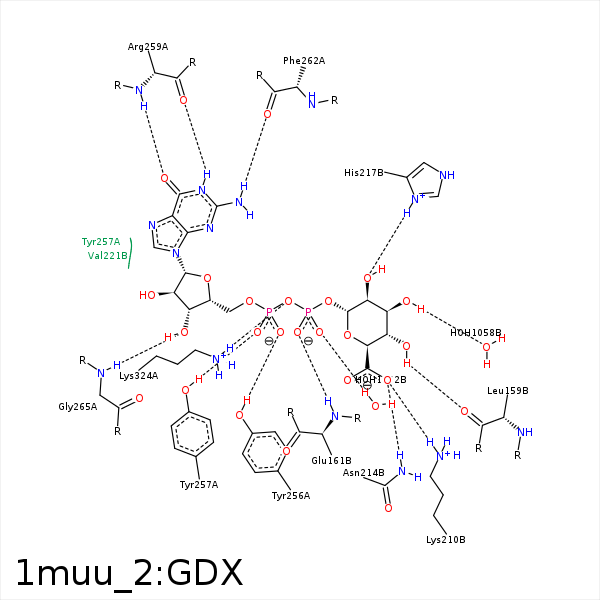


Represent the protein/ligand binding mode, centered on the ligand
Dashed lines represents hydrogen bonds and metal interactions
Green residue labels for amino acids with hydrophobic contacts (green lines) to the ligand
| Ligand | Protein | Interaction | |||
|---|---|---|---|---|---|
| Atom | Atom | Residue | Distance (Å) | Angle (°) | Type |
| O4' | O | PHE- 158 | 3.33 | 124.8 | H-Bond (Ligand Donor) |
| O4' | O | LEU- 159 | 2.82 | 135.7 | H-Bond (Ligand Donor) |
| C3' | CG | ARG- 160 | 3.79 | 0 | Hydrophobic |
| O2B | N | GLU- 161 | 2.92 | 165.9 | H-Bond (Protein Donor) |
| O6A | NZ | LYS- 210 | 2.97 | 160.36 | H-Bond (Protein Donor) |
| O6A | NZ | LYS- 210 | 2.97 | 0 | Ionic (Protein Cationic) |
| O6A | ND2 | ASN- 214 | 2.82 | 172.1 | H-Bond (Protein Donor) |
| O2A | OH | TYR- 256 | 2.73 | 155.69 | H-Bond (Protein Donor) |
| O1A | OH | TYR- 257 | 2.58 | 175.33 | H-Bond (Protein Donor) |
| N2 | O | ARG- 259 | 3.29 | 126.77 | H-Bond (Ligand Donor) |
| N1 | O | ARG- 259 | 2.58 | 167.48 | H-Bond (Ligand Donor) |
| O6 | N | ARG- 259 | 3.03 | 155.13 | H-Bond (Protein Donor) |
| N2 | O | PHE- 262 | 3.07 | 174.75 | H-Bond (Ligand Donor) |
| C1D | CB | PHE- 264 | 4.34 | 0 | Hydrophobic |
| C4D | CB | PHE- 264 | 3.88 | 0 | Hydrophobic |
| O3D | N | GLY- 265 | 2.92 | 151.13 | H-Bond (Protein Donor) |
| C5' | CB | CYS- 268 | 4.13 | 0 | Hydrophobic |
| C5D | CD2 | LEU- 269 | 3.81 | 0 | Hydrophobic |
| C1' | CD1 | LEU- 269 | 3.77 | 0 | Hydrophobic |
| C3D | CD2 | PHE- 323 | 3.82 | 0 | Hydrophobic |
| C5D | CE2 | PHE- 323 | 3.93 | 0 | Hydrophobic |
| O2A | NZ | LYS- 324 | 2.71 | 0 | Ionic (Protein Cationic) |
| O3A | NZ | LYS- 324 | 3.31 | 171.69 | H-Bond (Protein Donor) |
| O3B | O | HOH- 1032 | 2.7 | 179.96 | H-Bond (Protein Donor) |
| O3' | O | HOH- 1058 | 3.21 | 173.6 | H-Bond (Ligand Donor) |
