sc-PDB
An Annotated Database of Druggable Binding Sites from the Protein DataBank
An Annotated Database of Druggable Binding Sites from the Protein DataBank
1.630 Å
X-ray
2002-08-14
| Name: | Alcohol dehydrogenase |
|---|---|
| ID: | ADH_DROME |
| AC: | P00334 |
| Organism: | Drosophila melanogaster |
| Reign: | Eukaryota |
| TaxID: | 7227 |
| EC Number: | 1.1.1.1 |
| Chain Name: | Percentage of Residues within binding site |
|---|---|
| B | 100 % |
| B-Factor: | 12.377 |
|---|---|
| Number of residues: | 48 |
| Including | |
| Standard Amino Acids: | 45 |
| Non Standard Amino Acids: | 0 |
| Water Molecules: | 3 |
| Cofactors: | |
| Metals: | |
| Ligandability | Volume (Å3) |
|---|---|
| 1.149 | 597.375 |
| % Hydrophobic | % Polar |
|---|---|
| 50.85 | 49.15 |
| According to VolSite | |
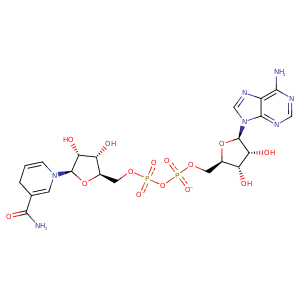
| HET Code: | NAI |
|---|---|
| Formula: | C21H27N7O14P2 |
| Molecular weight: | 663.425 g/mol |
| DrugBank ID: | DB00157 |
| Buried Surface Area: | 76.55 % |
| Polar Surface area: | 342.9 Å2 |
| Number of | |
|---|---|
| H-Bond Acceptors: | 19 |
| H-Bond Donors: | 6 |
| Rings: | 5 |
| Aromatic rings: | 2 |
| Anionic atoms: | 2 |
| Cationic atoms: | 0 |
| Rule of Five Violation: | 3 |
| Rotatable Bonds: | 11 |
| X | Y | Z |
|---|---|---|
| 21.1557 | 9.81882 | 9.34207 |
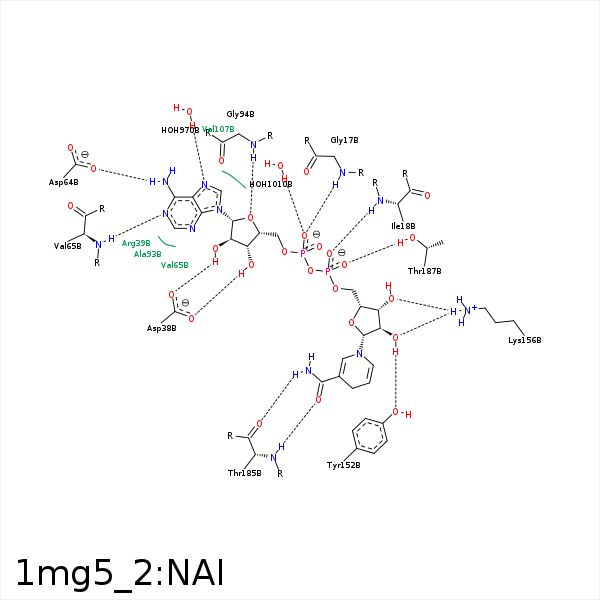


Represent the protein/ligand binding mode, centered on the ligand
Dashed lines represents hydrogen bonds and metal interactions
Green residue labels for amino acids with hydrophobic contacts (green lines) to the ligand
| Ligand | Protein | Interaction | |||
|---|---|---|---|---|---|
| Atom | Atom | Residue | Distance (Å) | Angle (°) | Type |
| C1B | CB | ALA- 13 | 3.47 | 0 | Hydrophobic |
| C4B | CB | ALA- 13 | 3.76 | 0 | Hydrophobic |
| O2A | N | GLY- 17 | 2.88 | 173.36 | H-Bond (Protein Donor) |
| O2N | N | ILE- 18 | 2.86 | 174.58 | H-Bond (Protein Donor) |
| C5D | CD1 | ILE- 18 | 4.37 | 0 | Hydrophobic |
| O3B | OD2 | ASP- 38 | 2.71 | 134.23 | H-Bond (Ligand Donor) |
| O3B | OD1 | ASP- 38 | 3.35 | 151.4 | H-Bond (Ligand Donor) |
| O2B | OD1 | ASP- 38 | 2.57 | 158.08 | H-Bond (Ligand Donor) |
| C3B | CD1 | ILE- 40 | 3.86 | 0 | Hydrophobic |
| N6A | OD1 | ASP- 64 | 2.99 | 164.48 | H-Bond (Ligand Donor) |
| N1A | N | VAL- 65 | 2.92 | 165.26 | H-Bond (Protein Donor) |
| C1B | CB | ALA- 93 | 4.31 | 0 | Hydrophobic |
| O4B | N | GLY- 94 | 3.27 | 162.22 | H-Bond (Protein Donor) |
| C4D | CG2 | ILE- 137 | 3.93 | 0 | Hydrophobic |
| C1D | CG2 | ILE- 137 | 3.75 | 0 | Hydrophobic |
| O2D | OH | TYR- 152 | 2.76 | 161.52 | H-Bond (Ligand Donor) |
| O3D | NZ | LYS- 156 | 3 | 148.99 | H-Bond (Protein Donor) |
| O2D | NZ | LYS- 156 | 3.04 | 131.2 | H-Bond (Protein Donor) |
| C4N | CB | PRO- 182 | 3.66 | 0 | Hydrophobic |
| O7N | N | THR- 185 | 2.82 | 162.12 | H-Bond (Protein Donor) |
| N7N | O | THR- 185 | 3.14 | 143.44 | H-Bond (Ligand Donor) |
| O1N | OG1 | THR- 187 | 2.57 | 156.43 | H-Bond (Protein Donor) |
| C2D | CD2 | LEU- 189 | 3.85 | 0 | Hydrophobic |
| N7A | O | HOH- 970 | 2.88 | 179.95 | H-Bond (Protein Donor) |
| O2A | O | HOH- 1010 | 2.62 | 179.98 | H-Bond (Protein Donor) |
