sc-PDB
An Annotated Database of Druggable Binding Sites from the Protein DataBank
An Annotated Database of Druggable Binding Sites from the Protein DataBank
1.900 Å
X-ray
2002-05-15
| Name: | UDP-glucose 4-epimerase |
|---|---|
| ID: | GALE_ECOLI |
| AC: | P09147 |
| Organism: | Escherichia coli |
| Reign: | Bacteria |
| TaxID: | 83333 |
| EC Number: | 5.1.3.2 |
| Chain Name: | Percentage of Residues within binding site |
|---|---|
| A | 100 % |
| B-Factor: | 28.631 |
|---|---|
| Number of residues: | 39 |
| Including | |
| Standard Amino Acids: | 38 |
| Non Standard Amino Acids: | 1 |
| Water Molecules: | 0 |
| Cofactors: | NAD |
| Metals: | |
| Ligandability | Volume (Å3) |
|---|---|
| 0.887 | 1069.875 |
| % Hydrophobic | % Polar |
|---|---|
| 40.38 | 59.62 |
| According to VolSite | |
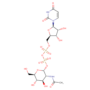
| HET Code: | UD1 |
|---|---|
| Formula: | C17H25N3O17P2 |
| Molecular weight: | 605.338 g/mol |
| DrugBank ID: | DB03397 |
| Buried Surface Area: | 71 % |
| Polar Surface area: | 325.69 Å2 |
| Number of | |
|---|---|
| H-Bond Acceptors: | 17 |
| H-Bond Donors: | 7 |
| Rings: | 3 |
| Aromatic rings: | 0 |
| Anionic atoms: | 2 |
| Cationic atoms: | 0 |
| Rule of Five Violation: | 3 |
| Rotatable Bonds: | 10 |
| X | Y | Z |
|---|---|---|
| 17.099 | 11.0771 | 37.1395 |
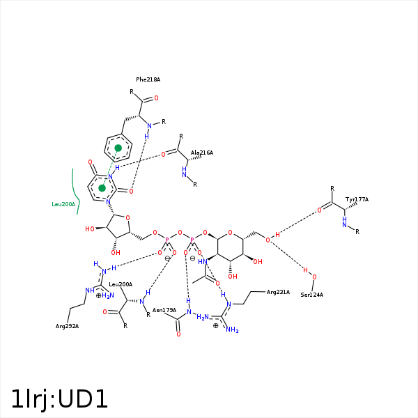


Represent the protein/ligand binding mode, centered on the ligand
Dashed lines represents hydrogen bonds and metal interactions
Green residue labels for amino acids with hydrophobic contacts (green lines) to the ligand
| Ligand | Protein | Interaction | |||
|---|---|---|---|---|---|
| Atom | Atom | Residue | Distance (Å) | Angle (°) | Type |
| O3' | O | LYS- 84 | 3.36 | 123.91 | H-Bond (Ligand Donor) |
| C3' | CG2 | VAL- 86 | 3.27 | 0 | Hydrophobic |
| O6' | OG | SER- 124 | 2.72 | 151.17 | H-Bond (Protein Donor) |
| O6' | O | TYR- 177 | 2.91 | 159.95 | H-Bond (Ligand Donor) |
| C6' | CD2 | PHE- 178 | 4.3 | 0 | Hydrophobic |
| O1B | ND2 | ASN- 179 | 3.22 | 160.36 | H-Bond (Protein Donor) |
| C1B | CD1 | LEU- 200 | 4.27 | 0 | Hydrophobic |
| C4B | CD2 | LEU- 200 | 4.35 | 0 | Hydrophobic |
| C5B | CB | LEU- 200 | 4.32 | 0 | Hydrophobic |
| O2A | N | LEU- 200 | 3.13 | 153.66 | H-Bond (Protein Donor) |
| N3 | O | ALA- 216 | 3.04 | 155 | H-Bond (Ligand Donor) |
| O2 | N | PHE- 218 | 3.32 | 164.72 | H-Bond (Protein Donor) |
| O2B | NE | ARG- 231 | 3.13 | 153.94 | H-Bond (Protein Donor) |
| C4B | CG | ARG- 231 | 3.76 | 0 | Hydrophobic |
| C5B | CZ | TYR- 233 | 4.26 | 0 | Hydrophobic |
| C1B | CG2 | VAL- 269 | 3.81 | 0 | Hydrophobic |
| C4B | CG2 | VAL- 269 | 3.8 | 0 | Hydrophobic |
| O1A | NH2 | ARG- 292 | 2.67 | 140.85 | H-Bond (Protein Donor) |
| O1A | CZ | ARG- 292 | 3.85 | 0 | Ionic (Protein Cationic) |
| C6' | C4N | NAD- 340 | 3.46 | 0 | Hydrophobic |
| C4' | C4N | NAD- 340 | 4.07 | 0 | Hydrophobic |
