sc-PDB
An Annotated Database of Druggable Binding Sites from the Protein DataBank
An Annotated Database of Druggable Binding Sites from the Protein DataBank
2.100 Å
X-ray
2001-10-23
| Name: | NAD(P)H-dependent D-xylose reductase |
|---|---|
| ID: | XYL1_CANTE |
| AC: | O74237 |
| Organism: | Candida tenuis |
| Reign: | Eukaryota |
| TaxID: | 45596 |
| EC Number: | 1.1.1.307 |
| Chain Name: | Percentage of Residues within binding site |
|---|---|
| B | 100 % |
| B-Factor: | 21.778 |
|---|---|
| Number of residues: | 45 |
| Including | |
| Standard Amino Acids: | 43 |
| Non Standard Amino Acids: | 0 |
| Water Molecules: | 2 |
| Cofactors: | |
| Metals: | |
| Ligandability | Volume (Å3) |
|---|---|
| 0.683 | 958.500 |
| % Hydrophobic | % Polar |
|---|---|
| 42.61 | 57.39 |
| According to VolSite | |
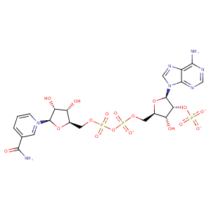
| HET Code: | NAP |
|---|---|
| Formula: | C21H25N7O17P3 |
| Molecular weight: | 740.381 g/mol |
| DrugBank ID: | DB03461 |
| Buried Surface Area: | 74.83 % |
| Polar Surface area: | 405.54 Å2 |
| Number of | |
|---|---|
| H-Bond Acceptors: | 21 |
| H-Bond Donors: | 5 |
| Rings: | 5 |
| Aromatic rings: | 3 |
| Anionic atoms: | 4 |
| Cationic atoms: | 1 |
| Rule of Five Violation: | 2 |
| Rotatable Bonds: | 13 |
| X | Y | Z |
|---|---|---|
| -1.16279 | 56.0537 | 50.5068 |


Represent the protein/ligand binding mode, centered on the ligand
Dashed lines represents hydrogen bonds and metal interactions
Green residue labels for amino acids with hydrophobic contacts (green lines) to the ligand
| Ligand | Protein | Interaction | |||
|---|---|---|---|---|---|
| Atom | Atom | Residue | Distance (Å) | Angle (°) | Type |
| O2D | N | CYS- 23 | 3.31 | 145.18 | H-Bond (Protein Donor) |
| O3D | N | TRP- 24 | 2.94 | 151.84 | H-Bond (Protein Donor) |
| C5N | CE3 | TRP- 24 | 3.36 | 0 | Hydrophobic |
| C3D | CB | TRP- 24 | 3.53 | 0 | Hydrophobic |
| O2D | OD2 | ASP- 47 | 2.68 | 171.25 | H-Bond (Ligand Donor) |
| C2D | CZ | TYR- 52 | 4.01 | 0 | Hydrophobic |
| N7N | OG | SER- 169 | 2.9 | 127.79 | H-Bond (Ligand Donor) |
| O7N | ND2 | ASN- 170 | 2.96 | 170.12 | H-Bond (Protein Donor) |
| N7N | OE1 | GLN- 191 | 3.04 | 144.17 | H-Bond (Ligand Donor) |
| C3N | CB | TYR- 217 | 4.19 | 0 | Hydrophobic |
| C5N | CB | TYR- 217 | 4.32 | 0 | Hydrophobic |
| DuAr | DuAr | TYR- 217 | 3.64 | 0 | Aromatic Face/Face |
| O2N | OG | SER- 218 | 3.06 | 158.01 | H-Bond (Protein Donor) |
| O5D | N | SER- 218 | 3.08 | 145.03 | H-Bond (Protein Donor) |
| O1A | N | PHE- 220 | 3.04 | 153.63 | H-Bond (Protein Donor) |
| C5B | CB | PHE- 220 | 3.95 | 0 | Hydrophobic |
| C4B | CD1 | PHE- 220 | 4.23 | 0 | Hydrophobic |
| C1B | CG | GLN- 223 | 4.09 | 0 | Hydrophobic |
| C4B | CG | GLN- 223 | 3.48 | 0 | Hydrophobic |
| O2N | OG | SER- 224 | 2.68 | 154.38 | H-Bond (Protein Donor) |
| C5D | CG1 | ILE- 272 | 4.43 | 0 | Hydrophobic |
| C4D | CD1 | ILE- 272 | 3.92 | 0 | Hydrophobic |
| C2D | CD1 | ILE- 272 | 4.44 | 0 | Hydrophobic |
| O2A | N | LYS- 274 | 3.02 | 161.31 | H-Bond (Protein Donor) |
| O2X | NZ | LYS- 274 | 2.68 | 169.17 | H-Bond (Protein Donor) |
| C5D | CB | LYS- 274 | 4.2 | 0 | Hydrophobic |
| C3D | CB | LYS- 274 | 4.44 | 0 | Hydrophobic |
| C3B | CD | LYS- 274 | 4.06 | 0 | Hydrophobic |
| O2X | NZ | LYS- 274 | 2.68 | 0 | Ionic (Protein Cationic) |
| O1X | OG | SER- 275 | 2.72 | 175.96 | H-Bond (Protein Donor) |
| O2X | N | ASN- 276 | 2.71 | 160.26 | H-Bond (Protein Donor) |
| O1X | CZ | ARG- 280 | 3.81 | 0 | Ionic (Protein Cationic) |
| O1X | NH1 | ARG- 280 | 2.75 | 156.56 | H-Bond (Protein Donor) |
| DuAr | CZ | ARG- 280 | 3.5 | 161.64 | Pi/Cation |
| N7A | ND2 | ASN- 284 | 2.93 | 166.79 | H-Bond (Protein Donor) |
| N6A | OD1 | ASN- 284 | 2.84 | 149.72 | H-Bond (Ligand Donor) |
| O3B | O | HOH- 2437 | 3.18 | 179.97 | H-Bond (Protein Donor) |
