sc-PDB
An Annotated Database of Druggable Binding Sites from the Protein DataBank
An Annotated Database of Druggable Binding Sites from the Protein DataBank
2.400 Å
X-ray
2001-08-29
| Name: | UDP-N-acetylhexosamine pyrophosphorylase |
|---|---|
| ID: | UAP1_HUMAN |
| AC: | Q16222 |
| Organism: | Homo sapiens |
| Reign: | Eukaryota |
| TaxID: | 9606 |
| EC Number: | 2.7.7.23 |
| Chain Name: | Percentage of Residues within binding site |
|---|---|
| A | 6 % |
| B | 94 % |
| B-Factor: | 33.492 |
|---|---|
| Number of residues: | 48 |
| Including | |
| Standard Amino Acids: | 47 |
| Non Standard Amino Acids: | 0 |
| Water Molecules: | 1 |
| Cofactors: | |
| Metals: | |
| Ligandability | Volume (Å3) |
|---|---|
| 0.421 | 928.125 |
| % Hydrophobic | % Polar |
|---|---|
| 34.91 | 65.09 |
| According to VolSite | |
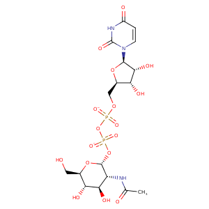
| HET Code: | UD1 |
|---|---|
| Formula: | C17H25N3O17P2 |
| Molecular weight: | 605.338 g/mol |
| DrugBank ID: | DB03397 |
| Buried Surface Area: | 69.9 % |
| Polar Surface area: | 325.69 Å2 |
| Number of | |
|---|---|
| H-Bond Acceptors: | 17 |
| H-Bond Donors: | 7 |
| Rings: | 3 |
| Aromatic rings: | 0 |
| Anionic atoms: | 2 |
| Cationic atoms: | 0 |
| Rule of Five Violation: | 3 |
| Rotatable Bonds: | 10 |
| X | Y | Z |
|---|---|---|
| -25.776 | 19.8408 | 22.828 |


Represent the protein/ligand binding mode, centered on the ligand
Dashed lines represents hydrogen bonds and metal interactions
Green residue labels for amino acids with hydrophobic contacts (green lines) to the ligand
| Ligand | Protein | Interaction | |||
|---|---|---|---|---|---|
| Atom | Atom | Residue | Distance (Å) | Angle (°) | Type |
| C1B | CB | LEU- 108 | 4.14 | 0 | Hydrophobic |
| O2' | O | LEU- 108 | 3.45 | 171.61 | H-Bond (Ligand Donor) |
| O2 | N | GLY- 110 | 2.82 | 135.1 | H-Bond (Protein Donor) |
| O2' | N | GLY- 111 | 3.09 | 134.61 | H-Bond (Protein Donor) |
| N3 | OE1 | GLN- 196 | 3.16 | 166.52 | H-Bond (Ligand Donor) |
| O4 | NE2 | GLN- 196 | 3.05 | 167.25 | H-Bond (Protein Donor) |
| O4 | N | GLY- 222 | 3.04 | 122.64 | H-Bond (Protein Donor) |
| O7' | ND2 | ASN- 223 | 2.7 | 154.35 | H-Bond (Protein Donor) |
| O4B | ND2 | ASN- 223 | 3.38 | 153.9 | H-Bond (Protein Donor) |
| C1B | CB | ASN- 223 | 4.22 | 0 | Hydrophobic |
| C3B | SG | CYS- 251 | 3.52 | 0 | Hydrophobic |
| C3B | CG1 | VAL- 252 | 4.12 | 0 | Hydrophobic |
| O3B | N | VAL- 252 | 3.02 | 166.56 | H-Bond (Protein Donor) |
| C6' | CG1 | VAL- 289 | 4.36 | 0 | Hydrophobic |
| O3' | N | GLY- 290 | 3.31 | 132.15 | H-Bond (Protein Donor) |
| O4' | N | GLY- 290 | 2.92 | 150.04 | H-Bond (Protein Donor) |
| N2' | OE2 | GLU- 303 | 2.74 | 171.29 | H-Bond (Ligand Donor) |
| O3' | OE1 | GLU- 303 | 2.59 | 153.74 | H-Bond (Ligand Donor) |
| C3' | CE2 | TYR- 304 | 4.4 | 0 | Hydrophobic |
| O1B | OH | TYR- 304 | 2.67 | 165.96 | H-Bond (Protein Donor) |
| C4' | CB | ASN- 327 | 4.32 | 0 | Hydrophobic |
| O3' | ND2 | ASN- 327 | 2.74 | 150.33 | H-Bond (Protein Donor) |
| O4' | O | ASN- 327 | 2.66 | 160.71 | H-Bond (Ligand Donor) |
| C8' | CD1 | PHE- 381 | 3.42 | 0 | Hydrophobic |
| C3' | CE2 | PHE- 383 | 4.5 | 0 | Hydrophobic |
| C8' | CE2 | PHE- 383 | 3.62 | 0 | Hydrophobic |
| C6' | CZ | PHE- 403 | 3.82 | 0 | Hydrophobic |
| O6' | NZ | LYS- 407 | 2.71 | 138.1 | H-Bond (Protein Donor) |
| O2B | NZ | LYS- 407 | 2.51 | 150.8 | H-Bond (Protein Donor) |
| O2B | NZ | LYS- 407 | 2.51 | 0 | Ionic (Protein Cationic) |
| O1A | NH2 | ARG- 453 | 2.83 | 164.51 | H-Bond (Protein Donor) |
| O1A | CZ | ARG- 453 | 3.71 | 0 | Ionic (Protein Cationic) |
| O1B | CZ | ARG- 453 | 3.65 | 0 | Ionic (Protein Cationic) |
| O6' | O | HOH- 974 | 3.19 | 179.98 | H-Bond (Protein Donor) |
