sc-PDB
An Annotated Database of Druggable Binding Sites from the Protein DataBank
An Annotated Database of Druggable Binding Sites from the Protein DataBank
2.200 Å
X-ray
2001-06-20
| Min | Mean | Median | Standard Deviation | Max | Count | |
|---|---|---|---|---|---|---|
| pChEMBL: | 5.700 | 5.700 | 5.700 | 0.000 | 5.700 | 2 |
| Name: | Tyrosine-protein phosphatase non-receptor type 1 |
|---|---|
| ID: | PTN1_HUMAN |
| AC: | P18031 |
| Organism: | Homo sapiens |
| Reign: | Eukaryota |
| TaxID: | 9606 |
| EC Number: | 3.1.3.48 |
| Chain Name: | Percentage of Residues within binding site |
|---|---|
| A | 19 % |
| B | 81 % |
| B-Factor: | 32.014 |
|---|---|
| Number of residues: | 33 |
| Including | |
| Standard Amino Acids: | 32 |
| Non Standard Amino Acids: | 0 |
| Water Molecules: | 1 |
| Cofactors: | |
| Metals: | |
| Ligandability | Volume (Å3) |
|---|---|
| 0.612 | 1836.000 |
| % Hydrophobic | % Polar |
|---|---|
| 38.05 | 61.95 |
| According to VolSite | |
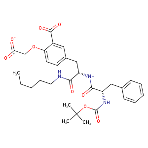
| HET Code: | TBH |
|---|---|
| Formula: | C31H46N3O9 |
| Molecular weight: | 604.712 g/mol |
| DrugBank ID: | DB02977 |
| Buried Surface Area: | 50.62 % |
| Polar Surface area: | 200.07 Å2 |
| Number of | |
|---|---|
| H-Bond Acceptors: | 11 |
| H-Bond Donors: | 6 |
| Rings: | 2 |
| Aromatic rings: | 2 |
| Anionic atoms: | 2 |
| Cationic atoms: | 1 |
| Rule of Five Violation: | 3 |
| Rotatable Bonds: | 21 |
| X | Y | Z |
|---|---|---|
| 19.1938 | 11.5927 | 42.1145 |
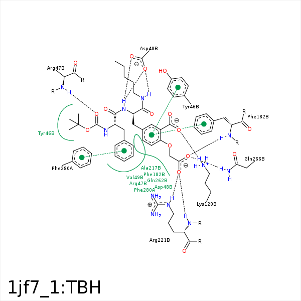


Represent the protein/ligand binding mode, centered on the ligand
Dashed lines represents hydrogen bonds and metal interactions
Green residue labels for amino acids with hydrophobic contacts (green lines) to the ligand
| Ligand | Protein | Interaction | |||
|---|---|---|---|---|---|
| Atom | Atom | Residue | Distance (Å) | Angle (°) | Type |
| C7 | CG | TYR- 46 | 3.75 | 0 | Hydrophobic |
| C39 | CD1 | TYR- 46 | 4.15 | 0 | Hydrophobic |
| O36 | N | ARG- 47 | 3.01 | 156.91 | H-Bond (Protein Donor) |
| C40 | CG | ARG- 47 | 4.41 | 0 | Hydrophobic |
| C31 | CD | ARG- 47 | 3.54 | 0 | Hydrophobic |
| N13 | OD2 | ASP- 48 | 3.04 | 133.39 | H-Bond (Ligand Donor) |
| N10 | OD2 | ASP- 48 | 2.89 | 138.05 | H-Bond (Ligand Donor) |
| N10 | OD1 | ASP- 48 | 2.8 | 153.24 | H-Bond (Ligand Donor) |
| C30 | CB | ASP- 48 | 3.86 | 0 | Hydrophobic |
| C7 | CG2 | VAL- 49 | 4.01 | 0 | Hydrophobic |
| O43 | NZ | LYS- 120 | 3.23 | 0 | Ionic (Protein Cationic) |
| O42 | NZ | LYS- 120 | 3.11 | 0 | Ionic (Protein Cationic) |
| O42 | NZ | LYS- 120 | 3.11 | 156.8 | H-Bond (Protein Donor) |
| C23 | CZ | PHE- 182 | 3.81 | 0 | Hydrophobic |
| C16 | CZ | PHE- 182 | 4.47 | 0 | Hydrophobic |
| O25 | N | PHE- 182 | 3.1 | 143.66 | H-Bond (Protein Donor) |
| C2 | CB | ALA- 217 | 3.35 | 0 | Hydrophobic |
| C2 | CG1 | ILE- 219 | 3.76 | 0 | Hydrophobic |
| C3 | CD1 | ILE- 219 | 3.88 | 0 | Hydrophobic |
| O25 | N | ARG- 221 | 3.47 | 125.66 | H-Bond (Protein Donor) |
| O26 | N | ARG- 221 | 3.05 | 173.28 | H-Bond (Protein Donor) |
| O26 | NE | ARG- 221 | 3.13 | 161.5 | H-Bond (Protein Donor) |
| C23 | CG | GLN- 262 | 4.06 | 0 | Hydrophobic |
| C2 | CG | GLN- 262 | 4.09 | 0 | Hydrophobic |
| O25 | NE2 | GLN- 266 | 2.95 | 151.14 | H-Bond (Protein Donor) |
| C28 | CZ | PHE- 280 | 4.09 | 0 | Hydrophobic |
