sc-PDB
An Annotated Database of Druggable Binding Sites from the Protein DataBank
An Annotated Database of Druggable Binding Sites from the Protein DataBank
2.400 Å
X-ray
2001-06-18
| Name: | Dihydrolipoyl dehydrogenase, mitochondrial |
|---|---|
| ID: | DLDH_YEAST |
| AC: | P09624 |
| Organism: | Saccharomyces cerevisiae |
| Reign: | Eukaryota |
| TaxID: | 559292 |
| EC Number: | 1.8.1.4 |
| Chain Name: | Percentage of Residues within binding site |
|---|---|
| A | 96 % |
| B | 4 % |
| B-Factor: | 22.372 |
|---|---|
| Number of residues: | 68 |
| Including | |
| Standard Amino Acids: | 68 |
| Non Standard Amino Acids: | 0 |
| Water Molecules: | 0 |
| Cofactors: | |
| Metals: | |
| Ligandability | Volume (Å3) |
|---|---|
| 1.302 | 1312.875 |
| % Hydrophobic | % Polar |
|---|---|
| 48.33 | 51.67 |
| According to VolSite | |
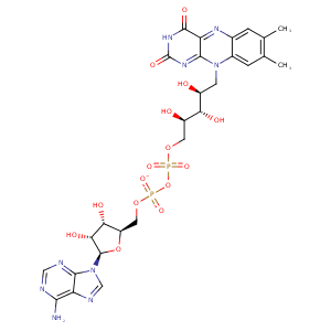
| HET Code: | FAD |
|---|---|
| Formula: | C27H31N9O15P2 |
| Molecular weight: | 783.534 g/mol |
| DrugBank ID: | DB03147 |
| Buried Surface Area: | 80.28 % |
| Polar Surface area: | 381.7 Å2 |
| Number of | |
|---|---|
| H-Bond Acceptors: | 22 |
| H-Bond Donors: | 7 |
| Rings: | 6 |
| Aromatic rings: | 3 |
| Anionic atoms: | 2 |
| Cationic atoms: | 0 |
| Rule of Five Violation: | 3 |
| Rotatable Bonds: | 13 |
| X | Y | Z |
|---|---|---|
| 44.9944 | 82.6579 | 12.9619 |
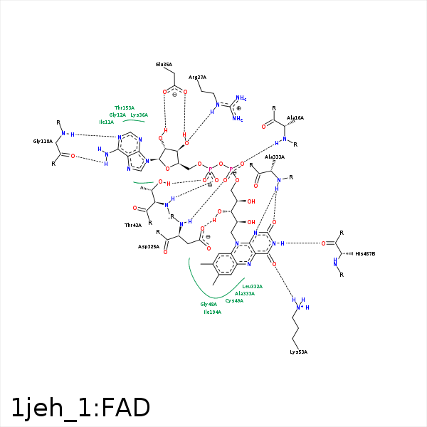


Represent the protein/ligand binding mode, centered on the ligand
Dashed lines represents hydrogen bonds and metal interactions
Green residue labels for amino acids with hydrophobic contacts (green lines) to the ligand
| Ligand | Protein | Interaction | |||
|---|---|---|---|---|---|
| Atom | Atom | Residue | Distance (Å) | Angle (°) | Type |
| C4' | CG | PRO- 15 | 4.04 | 0 | Hydrophobic |
| O1P | N | ALA- 16 | 2.96 | 158.95 | H-Bond (Protein Donor) |
| O3B | OE2 | GLU- 35 | 2.92 | 156.23 | H-Bond (Ligand Donor) |
| O3B | OE1 | GLU- 35 | 3.28 | 145.9 | H-Bond (Ligand Donor) |
| O2B | OE1 | GLU- 35 | 3 | 171.31 | H-Bond (Ligand Donor) |
| N3A | N | LYS- 36 | 3.44 | 143.7 | H-Bond (Protein Donor) |
| O3B | NE | ARG- 37 | 3.03 | 163.86 | H-Bond (Protein Donor) |
| O3B | NH2 | ARG- 37 | 3.38 | 140.76 | H-Bond (Protein Donor) |
| O1A | N | THR- 43 | 2.85 | 136.81 | H-Bond (Protein Donor) |
| O2A | OG1 | THR- 43 | 2.78 | 152.26 | H-Bond (Protein Donor) |
| C8M | CG2 | THR- 43 | 4.15 | 0 | Hydrophobic |
| C9 | CG2 | THR- 43 | 4.43 | 0 | Hydrophobic |
| C2' | CB | CYS- 44 | 4.2 | 0 | Hydrophobic |
| C4' | CB | CYS- 44 | 4.15 | 0 | Hydrophobic |
| O4' | N | CYS- 44 | 3.23 | 134.79 | H-Bond (Protein Donor) |
| C9A | SG | CYS- 49 | 4.41 | 0 | Hydrophobic |
| C2' | SG | CYS- 49 | 4.08 | 0 | Hydrophobic |
| C6 | CB | SER- 52 | 4.36 | 0 | Hydrophobic |
| C7M | CB | SER- 52 | 4.38 | 0 | Hydrophobic |
| O4 | NZ | LYS- 53 | 2.99 | 131.61 | H-Bond (Protein Donor) |
| N5 | NZ | LYS- 53 | 3.4 | 145.63 | H-Bond (Protein Donor) |
| N6A | O | GLY- 118 | 2.95 | 145.84 | H-Bond (Ligand Donor) |
| N1A | N | GLY- 118 | 2.93 | 156.45 | H-Bond (Protein Donor) |
| C7M | CB | SER- 173 | 3.96 | 0 | Hydrophobic |
| C8M | CB | SER- 173 | 4.31 | 0 | Hydrophobic |
| C7M | CD1 | LEU- 177 | 4.5 | 0 | Hydrophobic |
| C7M | CG2 | ILE- 194 | 3.63 | 0 | Hydrophobic |
| C8 | CD1 | ILE- 194 | 3.83 | 0 | Hydrophobic |
| C8M | CD | ARG- 285 | 3.93 | 0 | Hydrophobic |
| O3' | OD1 | ASP- 325 | 3.3 | 134.54 | H-Bond (Ligand Donor) |
| O3' | OD2 | ASP- 325 | 2.92 | 165.48 | H-Bond (Ligand Donor) |
| C5' | CB | ASP- 325 | 4.25 | 0 | Hydrophobic |
| O2P | N | ASP- 325 | 2.97 | 150.13 | H-Bond (Protein Donor) |
| N1 | N | ALA- 333 | 3.26 | 175.5 | H-Bond (Protein Donor) |
| O2 | N | ALA- 333 | 2.82 | 124.49 | H-Bond (Protein Donor) |
| C2' | CB | ALA- 333 | 4.18 | 0 | Hydrophobic |
| C4' | CB | ALA- 333 | 4.09 | 0 | Hydrophobic |
| C5' | CB | ALA- 336 | 3.98 | 0 | Hydrophobic |
| N3 | O | HIS- 457 | 2.75 | 153.24 | H-Bond (Ligand Donor) |
