sc-PDB
An Annotated Database of Druggable Binding Sites from the Protein DataBank
An Annotated Database of Druggable Binding Sites from the Protein DataBank
2.000 Å
X-ray
2001-03-08
| Name: | Cyclomaltodextrin glucanotransferase |
|---|---|
| ID: | CDGT_BACS0 |
| AC: | P05618 |
| Organism: | Bacillus sp. |
| Reign: | Bacteria |
| TaxID: | 1410 |
| EC Number: | 2.4.1.19 |
| Chain Name: | Percentage of Residues within binding site |
|---|---|
| A | 100 % |
| B-Factor: | 12.314 |
|---|---|
| Number of residues: | 20 |
| Including | |
| Standard Amino Acids: | 18 |
| Non Standard Amino Acids: | 0 |
| Water Molecules: | 2 |
| Cofactors: | |
| Metals: | |
| Ligandability | Volume (Å3) |
|---|---|
| 0.340 | 675.000 |
| % Hydrophobic | % Polar |
|---|---|
| 36.00 | 64.00 |
| According to VolSite | |
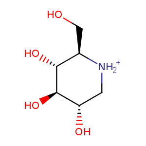
| HET Code: | NOJ |
|---|---|
| Formula: | C6H14NO4 |
| Molecular weight: | 164.180 g/mol |
| DrugBank ID: | DB03206 |
| Buried Surface Area: | 45.34 % |
| Polar Surface area: | 97.53 Å2 |
| Number of | |
|---|---|
| H-Bond Acceptors: | 4 |
| H-Bond Donors: | 5 |
| Rings: | 1 |
| Aromatic rings: | 0 |
| Anionic atoms: | 0 |
| Cationic atoms: | 1 |
| Rule of Five Violation: | 0 |
| Rotatable Bonds: | 1 |
| X | Y | Z |
|---|---|---|
| 45.2364 | 52.3488 | 19.2681 |
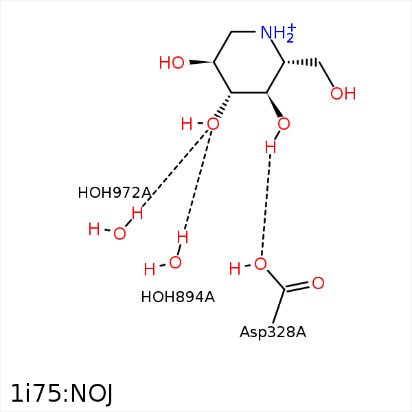


Represent the protein/ligand binding mode, centered on the ligand
Dashed lines represents hydrogen bonds and metal interactions
Green residue labels for amino acids with hydrophobic contacts (green lines) to the ligand
| Ligand | Protein | Interaction | |||
|---|---|---|---|---|---|
| Atom | Atom | Residue | Distance (Å) | Angle (°) | Type |
| C2 | CD1 | LEU- 197 | 4.45 | 0 | Hydrophobic |
| C6 | CB | ALA- 230 | 4.43 | 0 | Hydrophobic |
| O4 | OD2 | ASP- 328 | 3.12 | 132.07 | H-Bond (Ligand Donor) |
| O3 | O | HOH- 894 | 2.67 | 164.13 | H-Bond (Protein Donor) |
| O3 | O | HOH- 972 | 3.31 | 164.17 | H-Bond (Protein Donor) |
