sc-PDB
An Annotated Database of Druggable Binding Sites from the Protein DataBank
An Annotated Database of Druggable Binding Sites from the Protein DataBank
1.500 Å
X-ray
2001-02-15
| Name: | UDP-glucose 4-epimerase |
|---|---|
| ID: | GALE_HUMAN |
| AC: | Q14376 |
| Organism: | Homo sapiens |
| Reign: | Eukaryota |
| TaxID: | 9606 |
| EC Number: | / |
| Chain Name: | Percentage of Residues within binding site |
|---|---|
| A | 100 % |
| B-Factor: | 25.916 |
|---|---|
| Number of residues: | 43 |
| Including | |
| Standard Amino Acids: | 40 |
| Non Standard Amino Acids: | 1 |
| Water Molecules: | 2 |
| Cofactors: | NAD |
| Metals: | |
| Ligandability | Volume (Å3) |
|---|---|
| 1.097 | 1420.875 |
| % Hydrophobic | % Polar |
|---|---|
| 41.09 | 58.91 |
| According to VolSite | |
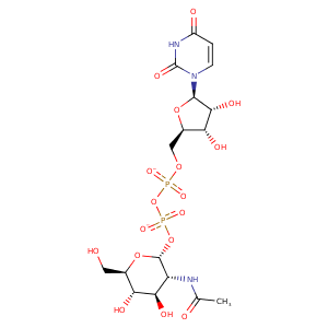
| HET Code: | UD1 |
|---|---|
| Formula: | C17H25N3O17P2 |
| Molecular weight: | 605.338 g/mol |
| DrugBank ID: | DB03397 |
| Buried Surface Area: | 57.93 % |
| Polar Surface area: | 325.69 Å2 |
| Number of | |
|---|---|
| H-Bond Acceptors: | 17 |
| H-Bond Donors: | 7 |
| Rings: | 3 |
| Aromatic rings: | 0 |
| Anionic atoms: | 2 |
| Cationic atoms: | 0 |
| Rule of Five Violation: | 3 |
| Rotatable Bonds: | 10 |
| X | Y | Z |
|---|---|---|
| 24.3425 | 18.3792 | 48.2029 |
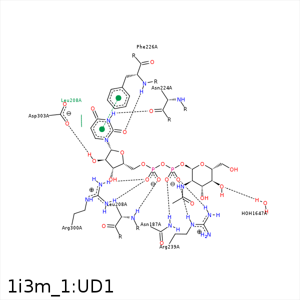


Represent the protein/ligand binding mode, centered on the ligand
Dashed lines represents hydrogen bonds and metal interactions
Green residue labels for amino acids with hydrophobic contacts (green lines) to the ligand
| Ligand | Protein | Interaction | |||
|---|---|---|---|---|---|
| Atom | Atom | Residue | Distance (Å) | Angle (°) | Type |
| C8' | CD | LYS- 92 | 4.5 | 0 | Hydrophobic |
| C1' | CG2 | THR- 134 | 4.35 | 0 | Hydrophobic |
| O1B | ND2 | ASN- 187 | 2.69 | 158.64 | H-Bond (Protein Donor) |
| C1B | CD1 | LEU- 208 | 4.47 | 0 | Hydrophobic |
| C4B | CD2 | LEU- 208 | 4.35 | 0 | Hydrophobic |
| C5B | CB | LEU- 208 | 4.06 | 0 | Hydrophobic |
| O2A | N | LEU- 208 | 2.94 | 175.36 | H-Bond (Protein Donor) |
| N3 | O | ASN- 224 | 2.89 | 176.84 | H-Bond (Ligand Donor) |
| O2 | N | PHE- 226 | 2.82 | 172.98 | H-Bond (Protein Donor) |
| C2B | CD1 | PHE- 226 | 4.13 | 0 | Hydrophobic |
| O1B | NE | ARG- 239 | 3.33 | 142.4 | H-Bond (Protein Donor) |
| O2B | NE | ARG- 239 | 3.12 | 153.66 | H-Bond (Protein Donor) |
| O2B | NH2 | ARG- 239 | 3.24 | 143.61 | H-Bond (Protein Donor) |
| O2B | CZ | ARG- 239 | 3.64 | 0 | Ionic (Protein Cationic) |
| C5B | CG | ARG- 239 | 3.96 | 0 | Hydrophobic |
| C5B | CZ | TYR- 241 | 4.49 | 0 | Hydrophobic |
| C1B | CG2 | VAL- 277 | 3.81 | 0 | Hydrophobic |
| C4B | CG2 | VAL- 277 | 4.13 | 0 | Hydrophobic |
| O5B | NH2 | ARG- 300 | 3.35 | 142.28 | H-Bond (Protein Donor) |
| O1A | NH2 | ARG- 300 | 2.87 | 146.61 | H-Bond (Protein Donor) |
| O1A | NH1 | ARG- 300 | 3.03 | 139.12 | H-Bond (Protein Donor) |
| O1A | CZ | ARG- 300 | 3.38 | 0 | Ionic (Protein Cationic) |
| O2' | OD2 | ASP- 303 | 2.63 | 146.31 | H-Bond (Ligand Donor) |
| C6' | CG1 | VAL- 304 | 4.38 | 0 | Hydrophobic |
| O4' | O | HOH- 1647 | 2.57 | 158.53 | H-Bond (Protein Donor) |
