sc-PDB
An Annotated Database of Druggable Binding Sites from the Protein DataBank
An Annotated Database of Druggable Binding Sites from the Protein DataBank
2.800 Å
X-ray
2001-01-10
| Name: | Glutamate dehydrogenase 1, mitochondrial |
|---|---|
| ID: | DHE3_BOVIN |
| AC: | P00366 |
| Organism: | Bos taurus |
| Reign: | Eukaryota |
| TaxID: | 9913 |
| EC Number: | 1.4.1.3 |
| Chain Name: | Percentage of Residues within binding site |
|---|---|
| D | 100 % |
| B-Factor: | 35.110 |
|---|---|
| Number of residues: | 49 |
| Including | |
| Standard Amino Acids: | 49 |
| Non Standard Amino Acids: | 0 |
| Water Molecules: | 0 |
| Cofactors: | |
| Metals: | |
| Ligandability | Volume (Å3) |
|---|---|
| 0.911 | 1076.625 |
| % Hydrophobic | % Polar |
|---|---|
| 32.60 | 67.40 |
| According to VolSite | |
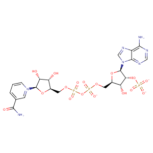
| HET Code: | NDP |
|---|---|
| Formula: | C21H26N7O17P3 |
| Molecular weight: | 741.389 g/mol |
| DrugBank ID: | DB02338 |
| Buried Surface Area: | 63.6 % |
| Polar Surface area: | 404.9 Å2 |
| Number of | |
|---|---|
| H-Bond Acceptors: | 22 |
| H-Bond Donors: | 5 |
| Rings: | 5 |
| Aromatic rings: | 2 |
| Anionic atoms: | 4 |
| Cationic atoms: | 0 |
| Rule of Five Violation: | 2 |
| Rotatable Bonds: | 13 |
| X | Y | Z |
|---|---|---|
| -25.2719 | 55.1482 | 61.8797 |
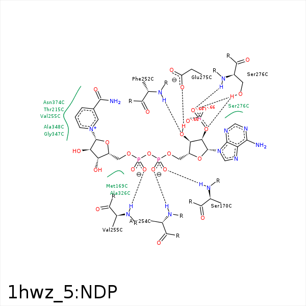


Represent the protein/ligand binding mode, centered on the ligand
Dashed lines represents hydrogen bonds and metal interactions
Green residue labels for amino acids with hydrophobic contacts (green lines) to the ligand
| Ligand | Protein | Interaction | |||
|---|---|---|---|---|---|
| Atom | Atom | Residue | Distance (Å) | Angle (°) | Type |
| O3X | NZ | LYS- 134 | 3.42 | 160 | H-Bond (Protein Donor) |
| O3X | NZ | LYS- 134 | 3.42 | 0 | Ionic (Protein Cationic) |
| C4D | CG | MET- 169 | 4.09 | 0 | Hydrophobic |
| C3D | CG | MET- 169 | 3.21 | 0 | Hydrophobic |
| O2A | N | SER- 170 | 3.19 | 148.35 | H-Bond (Protein Donor) |
| C4N | CG2 | THR- 215 | 3.75 | 0 | Hydrophobic |
| O3B | N | PHE- 252 | 3.23 | 145.03 | H-Bond (Protein Donor) |
| O1A | N | ASN- 254 | 2.81 | 160.21 | H-Bond (Protein Donor) |
| O2N | N | VAL- 255 | 3.33 | 176.26 | H-Bond (Protein Donor) |
| C5D | CG1 | VAL- 255 | 3.72 | 0 | Hydrophobic |
| C5N | CG1 | VAL- 255 | 3.73 | 0 | Hydrophobic |
| C3B | CG | GLU- 275 | 4.46 | 0 | Hydrophobic |
| O3B | OE1 | GLU- 275 | 3.09 | 172.36 | H-Bond (Ligand Donor) |
| O2B | N | SER- 276 | 3.21 | 134.21 | H-Bond (Protein Donor) |
| O2B | OG | SER- 276 | 2.7 | 156.09 | H-Bond (Protein Donor) |
| O2X | N | SER- 276 | 3.07 | 138.89 | H-Bond (Protein Donor) |
| O2X | OG | SER- 276 | 2.82 | 123.57 | H-Bond (Protein Donor) |
| O1X | NZ | LYS- 295 | 3.35 | 0 | Ionic (Protein Cationic) |
| C1B | CB | ALA- 326 | 3.98 | 0 | Hydrophobic |
| O3D | O | ALA- 326 | 3.45 | 135.71 | H-Bond (Ligand Donor) |
| N6A | OG | SER- 327 | 3.38 | 120.55 | H-Bond (Ligand Donor) |
| C4D | CB | ALA- 348 | 3.97 | 0 | Hydrophobic |
| O4D | N | ASN- 349 | 3.35 | 124.65 | H-Bond (Protein Donor) |
| C3N | CB | ASN- 374 | 3.81 | 0 | Hydrophobic |
| C3N | CA | GLU- 555 | 4.44 | 0 | Hydrophobic |
