sc-PDB
An Annotated Database of Druggable Binding Sites from the Protein DataBank
An Annotated Database of Druggable Binding Sites from the Protein DataBank
2.220 Å
X-ray
2001-01-09
| Name: | 3-hydroxy-3-methylglutaryl-coenzyme A reductase |
|---|---|
| ID: | HMDH_HUMAN |
| AC: | P04035 |
| Organism: | Homo sapiens |
| Reign: | Eukaryota |
| TaxID: | 9606 |
| EC Number: | 1.1.1.34 |
| Chain Name: | Percentage of Residues within binding site |
|---|---|
| B | 100 % |
| B-Factor: | 57.081 |
|---|---|
| Number of residues: | 24 |
| Including | |
| Standard Amino Acids: | 24 |
| Non Standard Amino Acids: | 0 |
| Water Molecules: | 0 |
| Cofactors: | |
| Metals: | |
| Ligandability | Volume (Å3) |
|---|---|
| 0.685 | 523.125 |
| % Hydrophobic | % Polar |
|---|---|
| 45.81 | 54.19 |
| According to VolSite | |
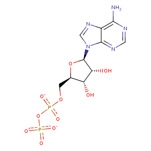
| HET Code: | ADP |
|---|---|
| Formula: | C10H12N5O10P2 |
| Molecular weight: | 424.177 g/mol |
| DrugBank ID: | - |
| Buried Surface Area: | 45.27 % |
| Polar Surface area: | 260.7 Å2 |
| Number of | |
|---|---|
| H-Bond Acceptors: | 14 |
| H-Bond Donors: | 3 |
| Rings: | 3 |
| Aromatic rings: | 2 |
| Anionic atoms: | 3 |
| Cationic atoms: | 0 |
| Rule of Five Violation: | 1 |
| Rotatable Bonds: | 6 |
| X | Y | Z |
|---|---|---|
| 19.7207 | -9.185 | -18.1292 |
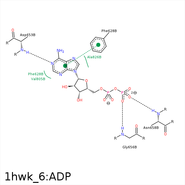


Represent the protein/ligand binding mode, centered on the ligand
Dashed lines represents hydrogen bonds and metal interactions
Green residue labels for amino acids with hydrophobic contacts (green lines) to the ligand
| Ligand | Protein | Interaction | |||
|---|---|---|---|---|---|
| Atom | Atom | Residue | Distance (Å) | Angle (°) | Type |
| C2' | CE1 | PHE- 628 | 4.49 | 0 | Hydrophobic |
| N6 | OD2 | ASP- 653 | 3.36 | 143.25 | H-Bond (Ligand Donor) |
| N1 | N | ASP- 653 | 2.7 | 157.73 | H-Bond (Protein Donor) |
| C5' | CB | ALA- 654 | 3.85 | 0 | Hydrophobic |
| O1B | N | GLY- 656 | 2.98 | 150.57 | H-Bond (Protein Donor) |
| O2B | N | ASN- 658 | 3.2 | 158.46 | H-Bond (Protein Donor) |
| C5' | CE | MET- 659 | 4.45 | 0 | Hydrophobic |
| C3' | CE | MET- 659 | 3.97 | 0 | Hydrophobic |
