sc-PDB
An Annotated Database of Druggable Binding Sites from the Protein DataBank
An Annotated Database of Druggable Binding Sites from the Protein DataBank
2.300 Å
X-ray
1995-01-02
| Name: | Guanine nucleotide-binding protein G(i) subunit alpha-1 |
|---|---|
| ID: | GNAI1_RAT |
| AC: | P10824 |
| Organism: | Rattus norvegicus |
| Reign: | Eukaryota |
| TaxID: | 10116 |
| EC Number: | / |
| Chain Name: | Percentage of Residues within binding site |
|---|---|
| A | 100 % |
| B-Factor: | 8.297 |
|---|---|
| Number of residues: | 46 |
| Including | |
| Standard Amino Acids: | 42 |
| Non Standard Amino Acids: | 1 |
| Water Molecules: | 3 |
| Cofactors: | |
| Metals: | MG |
| Ligandability | Volume (Å3) |
|---|---|
| 0.087 | 411.750 |
| % Hydrophobic | % Polar |
|---|---|
| 42.62 | 57.38 |
| According to VolSite | |
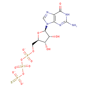
| HET Code: | GSP |
|---|---|
| Formula: | C10H14N5O13P3S |
| Molecular weight: | 537.230 g/mol |
| DrugBank ID: | DB01864 |
| Buried Surface Area: | 83.04 % |
| Polar Surface area: | 344.91 Å2 |
| Number of | |
|---|---|
| H-Bond Acceptors: | 17 |
| H-Bond Donors: | 6 |
| Rings: | 3 |
| Aromatic rings: | 1 |
| Anionic atoms: | 2 |
| Cationic atoms: | 0 |
| Rule of Five Violation: | 3 |
| Rotatable Bonds: | 8 |
| X | Y | Z |
|---|---|---|
| 38.881 | -27.6381 | 7.35497 |
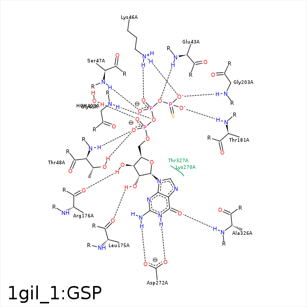


Represent the protein/ligand binding mode, centered on the ligand
Dashed lines represents hydrogen bonds and metal interactions
Green residue labels for amino acids with hydrophobic contacts (green lines) to the ligand
| Ligand | Protein | Interaction | |||
|---|---|---|---|---|---|
| Atom | Atom | Residue | Distance (Å) | Angle (°) | Type |
| O3B | N | GLU- 43 | 2.91 | 147.8 | H-Bond (Protein Donor) |
| C5' | CG | GLU- 43 | 4.04 | 0 | Hydrophobic |
| O1B | N | SER- 44 | 3.22 | 124.79 | H-Bond (Protein Donor) |
| O1B | N | GLY- 45 | 3.32 | 133.4 | H-Bond (Protein Donor) |
| O3A | N | GLY- 45 | 3.27 | 151.18 | H-Bond (Protein Donor) |
| O3G | NZ | LYS- 46 | 3.05 | 161.69 | H-Bond (Protein Donor) |
| O1B | N | LYS- 46 | 3.25 | 138.8 | H-Bond (Protein Donor) |
| O1B | NZ | LYS- 46 | 2.89 | 141.21 | H-Bond (Protein Donor) |
| O3G | NZ | LYS- 46 | 3.05 | 0 | Ionic (Protein Cationic) |
| O1B | NZ | LYS- 46 | 2.89 | 0 | Ionic (Protein Cationic) |
| O2B | N | SER- 47 | 2.82 | 154.96 | H-Bond (Protein Donor) |
| O1A | N | THR- 48 | 3.14 | 137.53 | H-Bond (Protein Donor) |
| O1A | OG1 | THR- 48 | 2.93 | 165.17 | H-Bond (Protein Donor) |
| C1' | CB | ASP- 150 | 4.26 | 0 | Hydrophobic |
| O3' | OG | SER- 151 | 3.2 | 132.11 | H-Bond (Ligand Donor) |
| O2' | O | LEU- 175 | 2.9 | 155.14 | H-Bond (Ligand Donor) |
| O3' | O | ARG- 176 | 2.85 | 122.62 | H-Bond (Ligand Donor) |
| C3' | CB | ARG- 178 | 3.85 | 0 | Hydrophobic |
| C4' | CG | ARG- 178 | 3.95 | 0 | Hydrophobic |
| O2G | N | THR- 181 | 3.17 | 149.82 | H-Bond (Protein Donor) |
| O3G | N | GLY- 203 | 2.69 | 156.03 | H-Bond (Protein Donor) |
| N7 | ND2 | ASN- 269 | 3.36 | 147.87 | H-Bond (Protein Donor) |
| O6 | N | LYS- 270 | 3.37 | 120.96 | H-Bond (Protein Donor) |
| N1 | OD1 | ASP- 272 | 2.86 | 155.28 | H-Bond (Ligand Donor) |
| N1 | OD2 | ASP- 272 | 3.48 | 139.89 | H-Bond (Ligand Donor) |
| N2 | OD2 | ASP- 272 | 3 | 167.13 | H-Bond (Ligand Donor) |
| O6 | N | ALA- 326 | 3.03 | 125.49 | H-Bond (Protein Donor) |
| O2G | MG | MG- 356 | 2.23 | 0 | Metal Acceptor |
| O2B | MG | MG- 356 | 2.35 | 0 | Metal Acceptor |
| O2A | O | HOH- 403 | 2.75 | 156.12 | H-Bond (Protein Donor) |
