sc-PDB
An Annotated Database of Druggable Binding Sites from the Protein DataBank
An Annotated Database of Druggable Binding Sites from the Protein DataBank
1.300 Å
X-ray
2000-04-13
| Name: | Dimethyl sulfoxide/trimethylamine N-oxide reductase |
|---|---|
| ID: | DSTOR_RHOSH |
| AC: | Q57366 |
| Organism: | Rhodobacter sphaeroides |
| Reign: | Bacteria |
| TaxID: | 1063 |
| EC Number: | / |
| Chain Name: | Percentage of Residues within binding site |
|---|---|
| A | 100 % |
| B-Factor: | 10.135 |
|---|---|
| Number of residues: | 58 |
| Including | |
| Standard Amino Acids: | 57 |
| Non Standard Amino Acids: | 1 |
| Water Molecules: | 0 |
| Cofactors: | |
| Metals: | |
| Ligandability | Volume (Å3) |
|---|---|
| 0.987 | 766.125 |
| % Hydrophobic | % Polar |
|---|---|
| 54.19 | 45.81 |
| According to VolSite | |
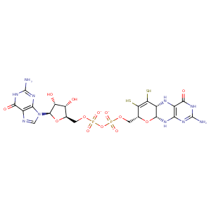
| HET Code: | MGD |
|---|---|
| Formula: | C20H24N10O13P2S2 |
| Molecular weight: | 738.541 g/mol |
| DrugBank ID: | - |
| Buried Surface Area: | 79.93 % |
| Polar Surface area: | 440.93 Å2 |
| Number of | |
|---|---|
| H-Bond Acceptors: | 22 |
| H-Bond Donors: | 10 |
| Rings: | 6 |
| Aromatic rings: | 1 |
| Anionic atoms: | 2 |
| Cationic atoms: | 0 |
| Rule of Five Violation: | 3 |
| Rotatable Bonds: | 9 |
| X | Y | Z |
|---|---|---|
| 22.2463 | 35.559 | 29.7791 |
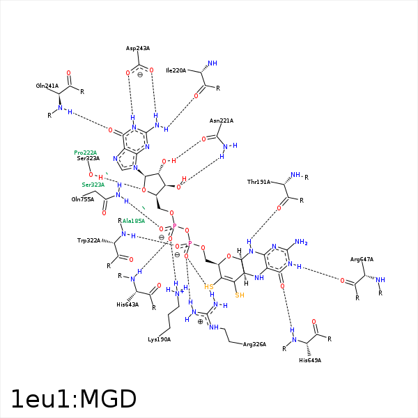


Represent the protein/ligand binding mode, centered on the ligand
Dashed lines represents hydrogen bonds and metal interactions
Green residue labels for amino acids with hydrophobic contacts (green lines) to the ligand
| Ligand | Protein | Interaction | |||
|---|---|---|---|---|---|
| Atom | Atom | Residue | Distance (Å) | Angle (°) | Type |
| S13 | CZ2 | TRP- 116 | 3.53 | 0 | Hydrophobic |
| S12 | CB | SER- 147 | 3.71 | 0 | Hydrophobic |
| O1B | NZ | LYS- 190 | 2.77 | 167.25 | H-Bond (Protein Donor) |
| O1B | NZ | LYS- 190 | 2.77 | 0 | Ionic (Protein Cationic) |
| N22 | O | THR- 191 | 2.96 | 155.7 | H-Bond (Ligand Donor) |
| C23 | CB | THR- 191 | 4.01 | 0 | Hydrophobic |
| C10 | CG2 | THR- 191 | 3.99 | 0 | Hydrophobic |
| N19 | OE2 | GLU- 193 | 3.48 | 126.15 | H-Bond (Ligand Donor) |
| N2 | O | ILE- 220 | 3.04 | 167.71 | H-Bond (Ligand Donor) |
| O3' | ND2 | ASN- 221 | 2.93 | 171.01 | H-Bond (Protein Donor) |
| O2' | OD1 | ASN- 221 | 2.72 | 163.82 | H-Bond (Ligand Donor) |
| O6 | N | GLN- 241 | 2.98 | 154.45 | H-Bond (Protein Donor) |
| N1 | OD2 | ASP- 243 | 2.81 | 149.85 | H-Bond (Ligand Donor) |
| N2 | OD1 | ASP- 243 | 2.86 | 151.06 | H-Bond (Ligand Donor) |
| O2A | N | TRP- 322 | 2.99 | 177.86 | H-Bond (Protein Donor) |
| S12 | CE3 | TRP- 322 | 3.45 | 0 | Hydrophobic |
| C5' | CB | SER- 323 | 4.14 | 0 | Hydrophobic |
| O4' | OG | SER- 323 | 2.89 | 171.82 | H-Bond (Protein Donor) |
| O2B | NH1 | ARG- 326 | 3.3 | 129.78 | H-Bond (Protein Donor) |
| O1A | NH2 | ARG- 326 | 2.81 | 148.36 | H-Bond (Protein Donor) |
| O1A | NH1 | ARG- 326 | 2.95 | 140.27 | H-Bond (Protein Donor) |
| O1A | CZ | ARG- 326 | 3.32 | 0 | Ionic (Protein Cationic) |
| C2' | CG1 | VAL- 640 | 4.21 | 0 | Hydrophobic |
| C2' | CB | SER- 642 | 4.44 | 0 | Hydrophobic |
| O1B | N | HIS- 643 | 3.12 | 163.76 | H-Bond (Protein Donor) |
| C10 | CB | HIS- 643 | 4.4 | 0 | Hydrophobic |
| N18 | O | ARG- 647 | 2.97 | 139.86 | H-Bond (Ligand Donor) |
| O17 | N | HIS- 649 | 2.94 | 150.05 | H-Bond (Protein Donor) |
| O2B | NE2 | GLN- 755 | 3 | 146.14 | H-Bond (Protein Donor) |
