sc-PDB
An Annotated Database of Druggable Binding Sites from the Protein DataBank
An Annotated Database of Druggable Binding Sites from the Protein DataBank
2.300 Å
X-ray
2000-08-15
| Name: | NADPH:adrenodoxin oxidoreductase, mitochondrial |
|---|---|
| ID: | ADRO_BOVIN |
| AC: | P08165 |
| Organism: | Bos taurus |
| Reign: | Eukaryota |
| TaxID: | 9913 |
| EC Number: | 1.18.1.6 |
| Chain Name: | Percentage of Residues within binding site |
|---|---|
| A | 98 % |
| B | 2 % |
| B-Factor: | 19.021 |
|---|---|
| Number of residues: | 62 |
| Including | |
| Standard Amino Acids: | 58 |
| Non Standard Amino Acids: | 0 |
| Water Molecules: | 4 |
| Cofactors: | |
| Metals: | |
| Ligandability | Volume (Å3) |
|---|---|
| 0.965 | 1674.000 |
| % Hydrophobic | % Polar |
|---|---|
| 49.19 | 50.81 |
| According to VolSite | |
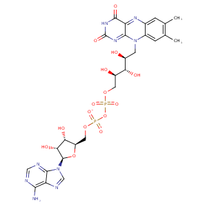
| HET Code: | FAD |
|---|---|
| Formula: | C27H31N9O15P2 |
| Molecular weight: | 783.534 g/mol |
| DrugBank ID: | DB03147 |
| Buried Surface Area: | 72.86 % |
| Polar Surface area: | 381.7 Å2 |
| Number of | |
|---|---|
| H-Bond Acceptors: | 22 |
| H-Bond Donors: | 7 |
| Rings: | 6 |
| Aromatic rings: | 3 |
| Anionic atoms: | 2 |
| Cationic atoms: | 0 |
| Rule of Five Violation: | 3 |
| Rotatable Bonds: | 13 |
| X | Y | Z |
|---|---|---|
| 7.86715 | 60.3763 | 123.169 |
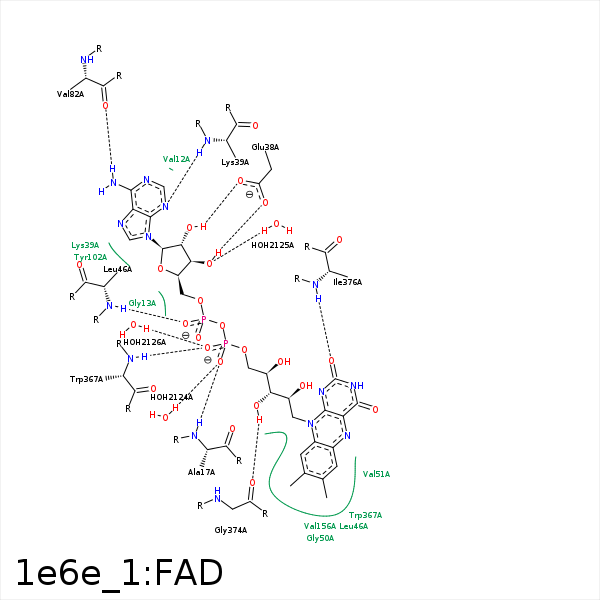


Represent the protein/ligand binding mode, centered on the ligand
Dashed lines represents hydrogen bonds and metal interactions
Green residue labels for amino acids with hydrophobic contacts (green lines) to the ligand
| Ligand | Protein | Interaction | |||
|---|---|---|---|---|---|
| Atom | Atom | Residue | Distance (Å) | Angle (°) | Type |
| C4' | CG | PRO- 16 | 3.93 | 0 | Hydrophobic |
| O1P | N | ALA- 17 | 2.72 | 146.11 | H-Bond (Protein Donor) |
| O3B | OE2 | GLU- 38 | 3.3 | 120.96 | H-Bond (Ligand Donor) |
| O3B | OE1 | GLU- 38 | 2.6 | 169.05 | H-Bond (Ligand Donor) |
| O2B | OE2 | GLU- 38 | 2.77 | 162.57 | H-Bond (Ligand Donor) |
| N3A | N | LYS- 39 | 3.11 | 145.04 | H-Bond (Protein Donor) |
| O2A | N | LEU- 46 | 3.09 | 171.25 | H-Bond (Protein Donor) |
| C8M | CD1 | LEU- 46 | 4.11 | 0 | Hydrophobic |
| C9 | CD1 | LEU- 46 | 4.34 | 0 | Hydrophobic |
| C2' | CB | LEU- 46 | 4.36 | 0 | Hydrophobic |
| C3' | CD1 | LEU- 46 | 4.38 | 0 | Hydrophobic |
| C4' | CB | LEU- 46 | 4.37 | 0 | Hydrophobic |
| N6A | O | VAL- 82 | 3.14 | 165.06 | H-Bond (Ligand Donor) |
| N1A | N | VAL- 82 | 3.16 | 132.17 | H-Bond (Protein Donor) |
| C1B | CB | TYR- 102 | 4.43 | 0 | Hydrophobic |
| C8M | CD | ARG- 124 | 3.57 | 0 | Hydrophobic |
| C7M | CG1 | VAL- 127 | 3.63 | 0 | Hydrophobic |
| C7M | CG1 | VAL- 156 | 3.76 | 0 | Hydrophobic |
| C8M | CG2 | VAL- 156 | 4.16 | 0 | Hydrophobic |
| C7 | CG2 | VAL- 156 | 3.91 | 0 | Hydrophobic |
| C8M | CE2 | TYR- 331 | 4.35 | 0 | Hydrophobic |
| C8M | CH2 | TRP- 367 | 4.02 | 0 | Hydrophobic |
| C5' | CE2 | TRP- 367 | 4.11 | 0 | Hydrophobic |
| C3' | CZ2 | TRP- 367 | 3.58 | 0 | Hydrophobic |
| O2P | N | TRP- 367 | 3.02 | 171.9 | H-Bond (Protein Donor) |
| O3' | O | GLY- 374 | 2.61 | 129.67 | H-Bond (Ligand Donor) |
| O2 | N | ILE- 376 | 2.83 | 172.55 | H-Bond (Protein Donor) |
| C2' | CG1 | ILE- 376 | 4.14 | 0 | Hydrophobic |
| O3' | OG1 | THR- 379 | 3.31 | 148.72 | H-Bond (Protein Donor) |
| C5' | CG2 | THR- 379 | 4.12 | 0 | Hydrophobic |
| O1P | O | HOH- 2124 | 2.61 | 160.89 | H-Bond (Protein Donor) |
| O3B | O | HOH- 2125 | 2.86 | 142.52 | H-Bond (Protein Donor) |
| O2P | O | HOH- 2126 | 2.79 | 179.96 | H-Bond (Protein Donor) |
