sc-PDB
An Annotated Database of Druggable Binding Sites from the Protein DataBank
An Annotated Database of Druggable Binding Sites from the Protein DataBank
2.100 Å
X-ray
2000-07-28
| Name: | Saccharopine dehydrogenase [NADP(+), L-glutamate-forming] |
|---|---|
| ID: | LYS9_MAGO7 |
| AC: | Q9P4R4 |
| Organism: | Magnaporthe oryzae |
| Reign: | Eukaryota |
| TaxID: | 242507 |
| EC Number: | 1.5.1.10 |
| Chain Name: | Percentage of Residues within binding site |
|---|---|
| A | 100 % |
| B-Factor: | 24.920 |
|---|---|
| Number of residues: | 49 |
| Including | |
| Standard Amino Acids: | 45 |
| Non Standard Amino Acids: | 1 |
| Water Molecules: | 3 |
| Cofactors: | |
| Metals: | |
| Ligandability | Volume (Å3) |
|---|---|
| 0.943 | 779.625 |
| % Hydrophobic | % Polar |
|---|---|
| 48.92 | 51.08 |
| According to VolSite | |
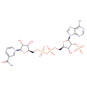
| HET Code: | NDP |
|---|---|
| Formula: | C21H26N7O17P3 |
| Molecular weight: | 741.389 g/mol |
| DrugBank ID: | DB02338 |
| Buried Surface Area: | 74.38 % |
| Polar Surface area: | 404.9 Å2 |
| Number of | |
|---|---|
| H-Bond Acceptors: | 22 |
| H-Bond Donors: | 5 |
| Rings: | 5 |
| Aromatic rings: | 2 |
| Anionic atoms: | 4 |
| Cationic atoms: | 0 |
| Rule of Five Violation: | 2 |
| Rotatable Bonds: | 13 |
| X | Y | Z |
|---|---|---|
| 37.9979 | 43.6141 | 68.7092 |


Represent the protein/ligand binding mode, centered on the ligand
Dashed lines represents hydrogen bonds and metal interactions
Green residue labels for amino acids with hydrophobic contacts (green lines) to the ligand
| Ligand | Protein | Interaction | |||
|---|---|---|---|---|---|
| Atom | Atom | Residue | Distance (Å) | Angle (°) | Type |
| O3B | N | SER- 11 | 3 | 154.85 | H-Bond (Protein Donor) |
| O1A | N | PHE- 13 | 3.05 | 159.77 | H-Bond (Protein Donor) |
| O2N | N | VAL- 14 | 3.18 | 175.85 | H-Bond (Protein Donor) |
| C5D | CG2 | VAL- 14 | 4.24 | 0 | Hydrophobic |
| C5N | CG1 | VAL- 14 | 3.89 | 0 | Hydrophobic |
| C2B | CB | CYS- 33 | 4.26 | 0 | Hydrophobic |
| O2X | NH2 | ARG- 34 | 3.34 | 156.39 | H-Bond (Protein Donor) |
| O3X | NE | ARG- 34 | 3.02 | 154.34 | H-Bond (Protein Donor) |
| O3X | CZ | ARG- 34 | 3.85 | 0 | Ionic (Protein Cationic) |
| O3X | N | THR- 35 | 3.26 | 138.71 | H-Bond (Protein Donor) |
| N6A | OD2 | ASP- 55 | 3.07 | 151.18 | H-Bond (Ligand Donor) |
| N1A | N | VAL- 56 | 3.12 | 169.94 | H-Bond (Protein Donor) |
| C5D | CB | LEU- 75 | 4.28 | 0 | Hydrophobic |
| C4B | CG2 | ILE- 76 | 4.4 | 0 | Hydrophobic |
| C1B | CG2 | ILE- 76 | 3.64 | 0 | Hydrophobic |
| O3D | O | ILE- 76 | 2.76 | 157.72 | H-Bond (Ligand Donor) |
| N7N | O | THR- 98 | 2.76 | 151.18 | H-Bond (Ligand Donor) |
| O7N | N | LEU- 125 | 2.87 | 136.69 | H-Bond (Protein Donor) |
| O7N | N | ASP- 126 | 2.72 | 171.88 | H-Bond (Protein Donor) |
| N7N | O | PRO- 127 | 3.01 | 157.54 | H-Bond (Ligand Donor) |
| C5N | CB | TRP- 174 | 3.67 | 0 | Hydrophobic |
| O1N | N | SER- 175 | 2.67 | 165.88 | H-Bond (Protein Donor) |
| C4N | SD | MET- 395 | 4.09 | 0 | Hydrophobic |
| C4N | CG2 | VAL- 399 | 3.31 | 0 | Hydrophobic |
| O2D | O1 | SHR- 501 | 2.68 | 152.47 | H-Bond (Ligand Donor) |
| C3N | C3 | SHR- 501 | 4.3 | 0 | Hydrophobic |
| C4N | C7 | SHR- 501 | 4.3 | 0 | Hydrophobic |
| O5B | O | HOH- 2062 | 3.11 | 156.51 | H-Bond (Protein Donor) |
