sc-PDB
An Annotated Database of Druggable Binding Sites from the Protein DataBank
An Annotated Database of Druggable Binding Sites from the Protein DataBank
2.500 Å
X-ray
1994-09-08
| Name: | Diphtheria toxin |
|---|---|
| ID: | DTX_CORBE |
| AC: | P00588 |
| Organism: | Corynephage beta |
| Reign: | Viruses |
| TaxID: | 10703 |
| EC Number: | / |
| Chain Name: | Percentage of Residues within binding site |
|---|---|
| A | 100 % |
| B-Factor: | 36.273 |
|---|---|
| Number of residues: | 34 |
| Including | |
| Standard Amino Acids: | 34 |
| Non Standard Amino Acids: | 0 |
| Water Molecules: | 0 |
| Cofactors: | |
| Metals: | |
| Ligandability | Volume (Å3) |
|---|---|
| 1.167 | 715.500 |
| % Hydrophobic | % Polar |
|---|---|
| 54.72 | 45.28 |
| According to VolSite | |
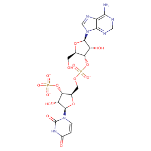
| HET Code: | APU |
|---|---|
| Formula: | C19H22N7O15P2 |
| Molecular weight: | 650.363 g/mol |
| DrugBank ID: | DB01792 |
| Buried Surface Area: | 56.35 % |
| Polar Surface area: | 348.81 Å2 |
| Number of | |
|---|---|
| H-Bond Acceptors: | 19 |
| H-Bond Donors: | 5 |
| Rings: | 5 |
| Aromatic rings: | 2 |
| Anionic atoms: | 3 |
| Cationic atoms: | 0 |
| Rule of Five Violation: | 2 |
| Rotatable Bonds: | 10 |
| X | Y | Z |
|---|---|---|
| 0.0135814 | 4.68921 | 47.7996 |
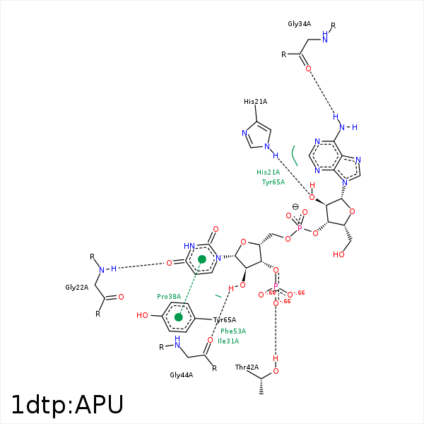


Represent the protein/ligand binding mode, centered on the ligand
Dashed lines represents hydrogen bonds and metal interactions
Green residue labels for amino acids with hydrophobic contacts (green lines) to the ligand
| Ligand | Protein | Interaction | |||
|---|---|---|---|---|---|
| Atom | Atom | Residue | Distance (Å) | Angle (°) | Type |
| O2B | NE2 | HIS- 21 | 3.09 | 161.71 | H-Bond (Protein Donor) |
| O4U | N | GLY- 22 | 2.99 | 142.61 | H-Bond (Protein Donor) |
| O1A | NZ | LYS- 24 | 3.67 | 0 | Ionic (Protein Cationic) |
| N6A | O | GLY- 34 | 2.92 | 167.48 | H-Bond (Ligand Donor) |
| C4B | CG | PRO- 38 | 4.48 | 0 | Hydrophobic |
| O2X | OG1 | THR- 42 | 3.33 | 164.82 | H-Bond (Protein Donor) |
| O2D | O | GLY- 44 | 3.12 | 160.86 | H-Bond (Ligand Donor) |
| C2D | CB | TYR- 54 | 4.39 | 0 | Hydrophobic |
| C1D | CE2 | TYR- 65 | 3.55 | 0 | Hydrophobic |
| C4D | CE2 | TYR- 65 | 4.24 | 0 | Hydrophobic |
| C5B | CZ2 | TRP- 153 | 4.48 | 0 | Hydrophobic |
