sc-PDB
An Annotated Database of Druggable Binding Sites from the Protein DataBank
An Annotated Database of Druggable Binding Sites from the Protein DataBank
2.310 Å
X-ray
1999-12-09
| Name: | UDP-glucose 6-dehydrogenase |
|---|---|
| ID: | UDG_STRPY |
| AC: | P0C0F4 |
| Organism: | Streptococcus pyogenes |
| Reign: | Bacteria |
| TaxID: | 1314 |
| EC Number: | / |
| Chain Name: | Percentage of Residues within binding site |
|---|---|
| A | 100 % |
| B-Factor: | 30.180 |
|---|---|
| Number of residues: | 48 |
| Including | |
| Standard Amino Acids: | 41 |
| Non Standard Amino Acids: | 1 |
| Water Molecules: | 6 |
| Cofactors: | NAD |
| Metals: | |
| Ligandability | Volume (Å3) |
|---|---|
| 0.243 | 621.000 |
| % Hydrophobic | % Polar |
|---|---|
| 42.39 | 57.61 |
| According to VolSite | |
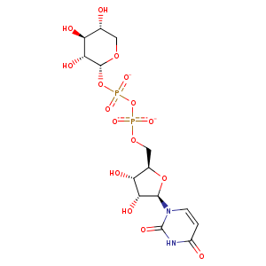
| HET Code: | UDX |
|---|---|
| Formula: | C14H20N2O16P2 |
| Molecular weight: | 534.260 g/mol |
| DrugBank ID: | DB01713 |
| Buried Surface Area: | 76.96 % |
| Polar Surface area: | 296.59 Å2 |
| Number of | |
|---|---|
| H-Bond Acceptors: | 16 |
| H-Bond Donors: | 6 |
| Rings: | 3 |
| Aromatic rings: | 0 |
| Anionic atoms: | 2 |
| Cationic atoms: | 0 |
| Rule of Five Violation: | 3 |
| Rotatable Bonds: | 8 |
| X | Y | Z |
|---|---|---|
| -13.6728 | 17.0686 | 54.4581 |
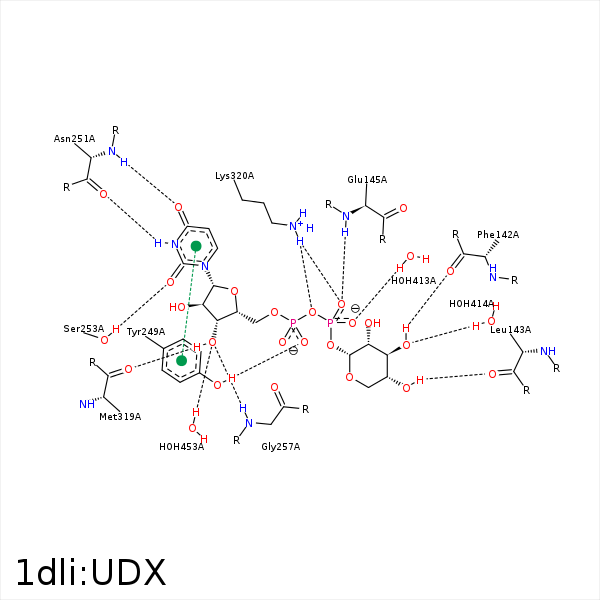


Represent the protein/ligand binding mode, centered on the ligand
Dashed lines represents hydrogen bonds and metal interactions
Green residue labels for amino acids with hydrophobic contacts (green lines) to the ligand
| Ligand | Protein | Interaction | |||
|---|---|---|---|---|---|
| Atom | Atom | Residue | Distance (Å) | Angle (°) | Type |
| O3' | O | PHE- 142 | 2.85 | 163.75 | H-Bond (Ligand Donor) |
| O4' | O | LEU- 143 | 2.83 | 160.44 | H-Bond (Ligand Donor) |
| C3' | CG | ARG- 144 | 3.86 | 0 | Hydrophobic |
| O2B | N | GLU- 145 | 2.91 | 162.68 | H-Bond (Protein Donor) |
| C2' | CD1 | LEU- 211 | 3.84 | 0 | Hydrophobic |
| C1D | CG2 | VAL- 215 | 4.23 | 0 | Hydrophobic |
| O2A | OH | TYR- 249 | 2.6 | 173.55 | H-Bond (Protein Donor) |
| O5D | OH | TYR- 249 | 3.42 | 123.09 | H-Bond (Protein Donor) |
| N3 | O | ASN- 251 | 2.84 | 162.91 | H-Bond (Ligand Donor) |
| O4 | N | ASN- 251 | 2.89 | 158.7 | H-Bond (Protein Donor) |
| O2 | OG | SER- 253 | 2.66 | 155.63 | H-Bond (Protein Donor) |
| C4D | CB | TYR- 256 | 4.3 | 0 | Hydrophobic |
| C1D | CB | TYR- 256 | 4.47 | 0 | Hydrophobic |
| O3D | N | GLY- 257 | 2.77 | 141.68 | H-Bond (Protein Donor) |
| C5' | SG | CYS- 260 | 3.82 | 0 | Hydrophobic |
| C1' | CD1 | LEU- 261 | 3.74 | 0 | Hydrophobic |
| C5D | CD2 | LEU- 261 | 3.66 | 0 | Hydrophobic |
| C3D | SD | MET- 319 | 4.08 | 0 | Hydrophobic |
| O3D | O | MET- 319 | 2.78 | 154.07 | H-Bond (Ligand Donor) |
| O2D | O | MET- 319 | 3.35 | 155.44 | H-Bond (Ligand Donor) |
| O2B | NZ | LYS- 320 | 3.03 | 146.46 | H-Bond (Protein Donor) |
| O3A | NZ | LYS- 320 | 3.25 | 147.73 | H-Bond (Protein Donor) |
| O2B | NZ | LYS- 320 | 3.03 | 0 | Ionic (Protein Cationic) |
| O1A | NZ | LYS- 320 | 3.67 | 0 | Ionic (Protein Cationic) |
| C2D | CB | LYS- 320 | 3.73 | 0 | Hydrophobic |
| C5' | C4N | NAD- 403 | 3.7 | 0 | Hydrophobic |
| O1B | O | HOH- 413 | 2.58 | 156.88 | H-Bond (Protein Donor) |
| O3' | O | HOH- 414 | 2.73 | 179.94 | H-Bond (Protein Donor) |
| O3D | O | HOH- 453 | 3.31 | 179.95 | H-Bond (Protein Donor) |
