sc-PDB
An Annotated Database of Druggable Binding Sites from the Protein DataBank
An Annotated Database of Druggable Binding Sites from the Protein DataBank
2.040 Å
X-ray
1999-09-29
| Min | Mean | Median | Standard Deviation | Max | Count | |
|---|---|---|---|---|---|---|
| pChEMBL: | 8.680 | 8.680 | 8.680 | 0.000 | 8.680 | 1 |
| Name: | Prothrombin |
|---|---|
| ID: | THRB_HUMAN |
| AC: | P00734 |
| Organism: | Homo sapiens |
| Reign: | Eukaryota |
| TaxID: | 9606 |
| EC Number: | 3.4.21.5 |
| Chain Name: | Percentage of Residues within binding site |
|---|---|
| B | 100 % |
| B-Factor: | 20.779 |
|---|---|
| Number of residues: | 35 |
| Including | |
| Standard Amino Acids: | 34 |
| Non Standard Amino Acids: | 0 |
| Water Molecules: | 1 |
| Cofactors: | |
| Metals: | |
| Ligandability | Volume (Å3) |
|---|---|
| 0.861 | 614.250 |
| % Hydrophobic | % Polar |
|---|---|
| 47.25 | 52.75 |
| According to VolSite | |
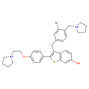
| HET Code: | BZT |
|---|---|
| Formula: | C32H37BrN2O2S |
| Molecular weight: | 593.617 g/mol |
| DrugBank ID: | - |
| Buried Surface Area: | 51.63 % |
| Polar Surface area: | 66.58 Å2 |
| Number of | |
|---|---|
| H-Bond Acceptors: | 2 |
| H-Bond Donors: | 3 |
| Rings: | 6 |
| Aromatic rings: | 4 |
| Anionic atoms: | 0 |
| Cationic atoms: | 2 |
| Rule of Five Violation: | 1 |
| Rotatable Bonds: | 9 |
| X | Y | Z |
|---|---|---|
| -19.2741 | -34.2671 | 20.3122 |
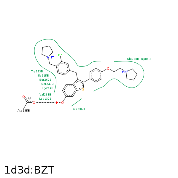


Represent the protein/ligand binding mode, centered on the ligand
Dashed lines represents hydrogen bonds and metal interactions
Green residue labels for amino acids with hydrophobic contacts (green lines) to the ligand
| Ligand | Protein | Interaction | |||
|---|---|---|---|---|---|
| Atom | Atom | Residue | Distance (Å) | Angle (°) | Type |
| BR1 | CB | HIS- 79 | 3.99 | 0 | Hydrophobic |
| BR1 | CZ | TYR- 83 | 3.55 | 0 | Hydrophobic |
| C24 | CH2 | TRP- 86 | 4.49 | 0 | Hydrophobic |
| BR1 | CZ3 | TRP- 86 | 4.16 | 0 | Hydrophobic |
| C29 | CE2 | TRP- 86 | 4.26 | 0 | Hydrophobic |
| C30 | CB | TRP- 86 | 3.82 | 0 | Hydrophobic |
| BR1 | CD1 | LEU- 132 | 3.83 | 0 | Hydrophobic |
| C23 | CG | LEU- 132 | 4.26 | 0 | Hydrophobic |
| C32 | CD1 | LEU- 132 | 4.23 | 0 | Hydrophobic |
| C17 | CD1 | ILE- 215 | 3.86 | 0 | Hydrophobic |
| O1 | OD1 | ASP- 235 | 2.55 | 145.63 | H-Bond (Ligand Donor) |
| C1 | CB | ALA- 236 | 4.07 | 0 | Hydrophobic |
| C20 | CB | GLU- 238 | 4.21 | 0 | Hydrophobic |
| C16 | CG | GLU- 238 | 3.6 | 0 | Hydrophobic |
| C7 | CB | SER- 241 | 3.61 | 0 | Hydrophobic |
| C4 | CB | SER- 241 | 4.37 | 0 | Hydrophobic |
| C6 | CG1 | VAL- 261 | 3.51 | 0 | Hydrophobic |
| C5 | CG1 | VAL- 261 | 3.62 | 0 | Hydrophobic |
| C23 | CD2 | TRP- 263 | 4.13 | 0 | Hydrophobic |
| C11 | CB | TRP- 263 | 4.21 | 0 | Hydrophobic |
| S1 | SG | CYS- 267 | 3.81 | 0 | Hydrophobic |
