sc-PDB
An Annotated Database of Druggable Binding Sites from the Protein DataBank
An Annotated Database of Druggable Binding Sites from the Protein DataBank
2.000 Å
X-ray
1999-07-22
| Name: | Methionine aminopeptidase |
|---|---|
| ID: | MAP1_ECOLI |
| AC: | P0AE18 |
| Organism: | Escherichia coli |
| Reign: | Bacteria |
| TaxID: | 83333 |
| EC Number: | / |
| Chain Name: | Percentage of Residues within binding site |
|---|---|
| A | 100 % |
| B-Factor: | 16.317 |
|---|---|
| Number of residues: | 23 |
| Including | |
| Standard Amino Acids: | 23 |
| Non Standard Amino Acids: | 0 |
| Water Molecules: | 0 |
| Cofactors: | |
| Metals: | |
| Ligandability | Volume (Å3) |
|---|---|
| 0.591 | 357.750 |
| % Hydrophobic | % Polar |
|---|---|
| 48.11 | 51.89 |
| According to VolSite | |

| HET Code: | MPH |
|---|---|
| Formula: | C4H11NO3PS |
| Molecular weight: | 184.174 g/mol |
| DrugBank ID: | DB02151 |
| Buried Surface Area: | 75.07 % |
| Polar Surface area: | 125.94 Å2 |
| Number of | |
|---|---|
| H-Bond Acceptors: | 4 |
| H-Bond Donors: | 1 |
| Rings: | 0 |
| Aromatic rings: | 0 |
| Anionic atoms: | 2 |
| Cationic atoms: | 1 |
| Rule of Five Violation: | 0 |
| Rotatable Bonds: | 4 |
| X | Y | Z |
|---|---|---|
| 21.54 | -13.8904 | 8.4722 |
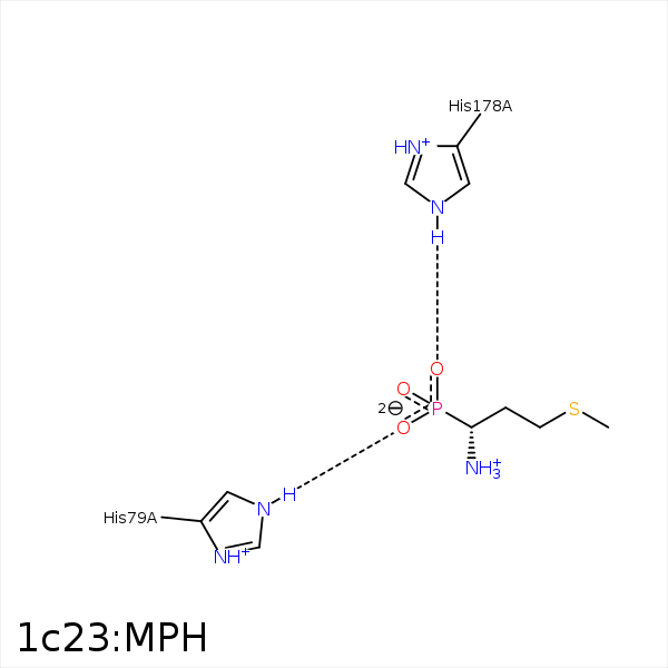


Represent the protein/ligand binding mode, centered on the ligand
Dashed lines represents hydrogen bonds and metal interactions
Green residue labels for amino acids with hydrophobic contacts (green lines) to the ligand
| Ligand | Protein | Interaction | |||
|---|---|---|---|---|---|
| Atom | Atom | Residue | Distance (Å) | Angle (°) | Type |
| CE | SG | CYS- 59 | 4.35 | 0 | Hydrophobic |
| CG | SG | CYS- 59 | 3.92 | 0 | Hydrophobic |
| SD | CD2 | TYR- 62 | 4 | 0 | Hydrophobic |
| CE | CD1 | TYR- 65 | 3.5 | 0 | Hydrophobic |
| CG | SG | CYS- 70 | 3.78 | 0 | Hydrophobic |
| CE | SG | CYS- 70 | 3.8 | 0 | Hydrophobic |
| O3 | NE2 | HIS- 79 | 3 | 159.67 | H-Bond (Protein Donor) |
| SD | CE2 | PHE- 177 | 3.92 | 0 | Hydrophobic |
| CB | CZ | PHE- 177 | 3.5 | 0 | Hydrophobic |
| CE | CZ3 | TRP- 221 | 3.72 | 0 | Hydrophobic |
