sc-PDB
An Annotated Database of Druggable Binding Sites from the Protein DataBank
An Annotated Database of Druggable Binding Sites from the Protein DataBank
2.000 Å
X-ray
1991-11-08
| Name: | Adenylate kinase |
|---|---|
| ID: | KAD_ECOLI |
| AC: | P69441 |
| Organism: | Escherichia coli |
| Reign: | Bacteria |
| TaxID: | 83333 |
| EC Number: | / |
| Chain Name: | Percentage of Residues within binding site |
|---|---|
| A | 100 % |
| B-Factor: | 22.566 |
|---|---|
| Number of residues: | 44 |
| Including | |
| Standard Amino Acids: | 42 |
| Non Standard Amino Acids: | 0 |
| Water Molecules: | 2 |
| Cofactors: | |
| Metals: | |
| Ligandability | Volume (Å3) |
|---|---|
| 0.112 | 347.625 |
| % Hydrophobic | % Polar |
|---|---|
| 50.49 | 49.51 |
| According to VolSite | |
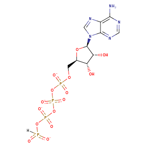
| HET Code: | AP5 |
|---|---|
| Formula: | C20H24N10O22P5 |
| Molecular weight: | 911.327 g/mol |
| DrugBank ID: | DB01717 |
| Buried Surface Area: | 68.79 % |
| Polar Surface area: | 543.69 Å2 |
| Number of | |
|---|---|
| H-Bond Acceptors: | 30 |
| H-Bond Donors: | 6 |
| Rings: | 6 |
| Aromatic rings: | 4 |
| Anionic atoms: | 5 |
| Cationic atoms: | 0 |
| Rule of Five Violation: | 3 |
| Rotatable Bonds: | 16 |
| X | Y | Z |
|---|---|---|
| 16.5798 | 46.3766 | 20.0894 |
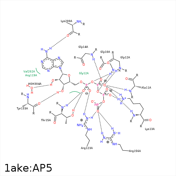


Represent the protein/ligand binding mode, centered on the ligand
Dashed lines represents hydrogen bonds and metal interactions
Green residue labels for amino acids with hydrophobic contacts (green lines) to the ligand
| Ligand | Protein | Interaction | |||
|---|---|---|---|---|---|
| Atom | Atom | Residue | Distance (Å) | Angle (°) | Type |
| O3B | N | GLY- 10 | 3.01 | 143.21 | H-Bond (Protein Donor) |
| O1G | N | GLY- 10 | 3.42 | 142.88 | H-Bond (Protein Donor) |
| O1B | N | ALA- 11 | 3.05 | 123.27 | H-Bond (Protein Donor) |
| O3A | N | GLY- 12 | 2.98 | 137.18 | H-Bond (Protein Donor) |
| O1B | N | GLY- 12 | 3.11 | 131.05 | H-Bond (Protein Donor) |
| O3A | N | LYS- 13 | 3.38 | 124.29 | H-Bond (Protein Donor) |
| O1B | NZ | LYS- 13 | 2.78 | 152.86 | H-Bond (Protein Donor) |
| O1B | N | LYS- 13 | 2.91 | 169.21 | H-Bond (Protein Donor) |
| O2G | NZ | LYS- 13 | 3.01 | 137.39 | H-Bond (Protein Donor) |
| O1B | NZ | LYS- 13 | 2.78 | 0 | Ionic (Protein Cationic) |
| O2G | NZ | LYS- 13 | 3.01 | 0 | Ionic (Protein Cationic) |
| O2B | N | GLY- 14 | 2.7 | 163.57 | H-Bond (Protein Donor) |
| O1A | N | THR- 15 | 2.85 | 150.3 | H-Bond (Protein Donor) |
| O1A | OG1 | THR- 15 | 2.71 | 159.08 | H-Bond (Protein Donor) |
| O1D | CZ | ARG- 36 | 3.72 | 0 | Ionic (Protein Cationic) |
| C4F | CB | ARG- 119 | 4.12 | 0 | Hydrophobic |
| DuAr | CZ | ARG- 119 | 3.68 | 3.64 | Pi/Cation |
| O2A | NH1 | ARG- 123 | 2.91 | 149.72 | H-Bond (Protein Donor) |
| O3B | NH1 | ARG- 123 | 3.27 | 137.35 | H-Bond (Protein Donor) |
| O3G | NH1 | ARG- 123 | 3.21 | 139.24 | H-Bond (Protein Donor) |
| O2A | CZ | ARG- 123 | 3.99 | 0 | Ionic (Protein Cationic) |
| O2D | CZ | ARG- 123 | 3.33 | 0 | Ionic (Protein Cationic) |
| C3F | CD | ARG- 123 | 3.8 | 0 | Hydrophobic |
| C4F | CD | ARG- 123 | 4.24 | 0 | Hydrophobic |
| C3F | CG2 | VAL- 132 | 3.7 | 0 | Hydrophobic |
| O3F | O | TYR- 133 | 2.96 | 159.96 | H-Bond (Ligand Donor) |
| C2F | CB | HIS- 134 | 4.13 | 0 | Hydrophobic |
| O1D | CZ | ARG- 156 | 3.18 | 0 | Ionic (Protein Cationic) |
| O2D | CZ | ARG- 156 | 2.55 | 0 | Ionic (Protein Cationic) |
| N6A | O | LYS- 200 | 2.77 | 170.71 | H-Bond (Ligand Donor) |
| O2F | O | HOH- 304 | 2.66 | 174.31 | H-Bond (Ligand Donor) |
